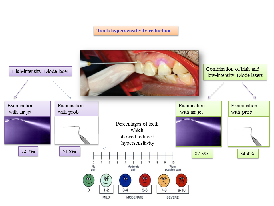
Logo-joddd

Visual Abstract

JODDD now with visual abstracts. Authors can now add a visual abstract to their submission.

Platinum* Open Access

*This Platinum Open Access journal publishes articles totally free of charge for the authors and provides unrestricted access to the published content through its website and open access repositories such as PubMed Central.

  Indexing & Abstracting

Following


Social media

J Dent Res Dent Clin Dent Prospects. 2017;11(2): 90-95. doi: 10.15171/joddd.2017.017
PMID: 28748049        PMCID: PMC5519999

Original Article

Effect of different nickel-titanium rotary files on dentinal crack formation during retreatment procedure

Mehmet Çitak 1, Taha Özyürek 2 *

Cited by CrossRef: 11


1- Bal S, Pare S, Unnikrishnan A, Shetty D, Kumar H, Ragher M. Detection of dentinal microcracks in radicular dentin after shaping with XP-endo Shaper, Neoendo flex files, and Hero Shaper using scanning electron microscope: An in vitro study. J Pharm Bioall Sci. 2020;12(5):259 [Crossref]
2- Das M, Shivakumar S, Das A, Mailankote S, Naik S, Sathydevi P. Assessment of root dentin defect during retreatment procedure using various NiTi hand and rotary retreatment files: An In vitro study. J Pharm Bioall Sci. 2022;14(5):573 [Crossref]
3- KONYALI İ, KOÇAK M, SAĞLAM B, TAZEGÜL-KOCAK S. NİKEL TİTANYUM EĞELERİN TEKRARLAYAN KÖK KANAL TEDAVİSİ SIRASINDA APİKAL DEBRİS TAŞKINLIĞININ KARŞILAŞTIRILMASI. J Int Dent Sci. 2023;9(1):19 [Crossref]
4- Zarean P, Özcan M, Zarean P, Haghani S, Jahromi M, Al-Haj Husain N, Khabiri M. Micro-Computed Tomographic Assessment of Microcrack Formation before and after Instrumentation of Curved Root Canals with Neoniti Rotary Files. Materials. 2022;15(9):3002 [Crossref]
5- Almeida A, Romeiro K, Cassimiro M, Gominho L, Dantas E, Silva S, Albuquerque D. Micro-CT analysis of dentinal microcracks on root canals filled with a bioceramic sealer and retreated with reciprocating instruments. Sci Rep. 2020;10(1) [Crossref]
6- Tejaswi S, Singh A, Manglekar S, Ambikathanaya U, Shetty S. Evaluation of dentinal crack propagation, amount of gutta percha remaining and time required during removal of gutta percha using two different rotary instruments and hand instruments - An In vitro study. Niger J Clin Pract. 2022;25(4):524 [Crossref]
7- AbuMostafa A, Almoqayyad H, Mohammad A, Testarelli L. A Digital Microscopic Inspection of Dentinal Defects after Using Endodontic Retreatment Files. International Journal of Dentistry. 2021;2021:1 [Crossref]
8- Boonchoo K, Leelataweewud P, Yanpiset K, Jirarattanasopha V. Simplify pulpectomy in primary molars with a single-file reciprocating system: a randomized controlled clinical trial. Clin Oral Invest. 2020;24(8):2683 [Crossref]
9- Tonelli S, Antunes M, Soares de Toubes K, Miranda A, Pereira A, Nunes E, Silveira F. Dentinal Microcracks Induced by Endodontic Procedures: Suggested Design for Experimental Studies Using Micro–Computed Tomography and Strain Gauges. Journal of Endodontics. 2021;47(7):1157 [Crossref]
10- Yüksel B, Öncü A, Çelİkten B, Bİlecenoğlu B, Orhan A, Orhan K. Micro‐CT evaluation of ‘danger zone’ and microcrack formation in mesial root canals of primary teeth with single‐file rotary and reciprocating systems. Int J Paed Dentistry. 2022;32(1):109 [Crossref]
11- Barakat R, Almohareb R, Alsayyar A, Almalki F, Alharbi H, Vivona M. Evaluation of Dentinal Microcracks following Diode Laser- and Ultrasonic-Activated Removal of Bioceramic Material during Root Canal Retreatment. Scanning. 2022;2022:1 [Crossref]