Push-out bond strength of intra-orifice barrier materials: Bulk-fill composite versus calcium silicate cement
J Dent Res Dent Clin Dent Prospects, 12(1), 6-11; DOI:10.15171/joddd.2018.002
Basic Research
Push-out bond strength of intra-orifice barrier materials: Bulk-fill composite versus calcium silicate cement
Taha Özyurek1, Gülşah Uslu2, Koray Yilmaz3*
1
Ondokuz Mayis University, Faculty of Dentistry, Department of Endodontics, Samsun, Turkey
2
Çorum Oral and Dental Health Hospital Çorum, Turkey
*Corresponding Author; Email: koray1903@hotmail.com
© 2017 by Tabriz University of Medical Sciences
Abstract
Background.The aim of this study was to compare the push-out bond strengths of calcium silicate-based ProRoot MTA and Biodentine cements and SureFil SDR and EverX Posterior bulk-fill composite resins.
Methods.
Twenty-four single-rooted maxillary central incisors were sectioned below the cementoenamel junction, and the root canals were instrumented using rotary files. Thereafter, a parallel post drill was used to obtain a standardized root canal dimension. The roots were randomly assigned to one of the following groups with respect to the intra-orifice barrier used: ProRoot MTA; Biodentine; SureFil SDR; EverX Posterior. Five 1-mm-thick sections were obtained from the coronal aspect of each root. Push-out bond strength testing was performed and data were analyzed with Kruskal-Wallis and post hoc Dunn tests (P<0.05).
Results.
SureFil SDR and EverX Posterior bulk-fill composite resins’ bond strengths were significantly higher than ProRoot MTA and Biodentine calcium silicate cements. However, no statistically significant differences were observed between bulk-fill composite resins values and calcium silicate cement values.
Conclusion.
Within the limitations of present study, calcium silicate-based ProRoot MTA cement’s push-out bond strength was lower than those of Biodentine, SureFil SDR and EverX Posterior materials.
Keywords: Bulk-fill composite, calcium-silicate, push-out
Introduction
Coronal leakage is one of the most important reasons for the failure after root canal treatment.1 Ray and Trope2 reported that the quality of coronal restoration is more important in protecting the periapical health than the quality of root canal filling. For this purpose, it has been suggested that to prevent the penetration of oral fluids and microorganisms into the root canals 3‒4 mm of coronal gutta-percha should be removed from the root canal and an intra-orifice barrier should be placed at canal orifice3 or a pulpal base should be placed using a restorative material.4 Previous studies have reported that covering the pulpal base with intra-orifice barrier materials after the root canal treatment constructs a secondary defense line against bacterial leakage.4,5 For this purpose, different materials have been employed, including temporary filling materials, glass-ionomer cement, composite resin, MTA and IRM.6,7
Today, MTA is also safely used in conservative pulpal treatments, root resorption treatments, and apexification procedures.8,9 MTA has been shown to be a bioactive material inducing the formation of hard tissues; it is well-tolerated by the tissues it contacts because of its biocompatibili.y10,11 However, long setting time of MTA (4‒6 hours) and the difficulties in adjusting the consistency while mixing make this material non-practical for clinical use.12 Numerous materials based on tri-calcium silicate have been developed and introduced to the market in order to eliminate these disadvantages of MTA.13-14 Biodentine (BD; Septodont, Saint-Maur-des-Fosses, France) is a new endodontic cement containing tri-calcium silicate and calcium carbonate, with a setting time of 12 minutes.15 The manufacturer claims that BD can be used as a replacement for dentin tissue for restorative purposes and as direct pulp cupping material for endodontic purposes, as well as restoration of perforations and as a root-end filling material.16
SureFil SDR flow (SDR; Dentsply Caulk, Milford, DE, USA), one of the bulk-fill composite resins recently introduced to the market, is a silorane-based nano- and micro-hybrid composite with low viscosity; its shrinkage stress is lower than conventional fluids.17,18 Another fiber-reinforced bulk-fill composite resin, also newly introduced to the market, is EverX Posterior (EXP; GC Dental Products Corp., Tokyo, Japan).
In comprehensive literature research, no study was found, comparing the push-out bond strengths of SDR and EXP bulk-fill composite resins. For this reason, the aim of the present study was to compare the push-out bond strengths of calcium silicate-based ProRoot MTA and BD cements and SDR and EXP bulk-fill composite resins. The null hypothesis of the present study was there would be no statistically significant difference between the push-out bond strengths of the tested materials.
Methods
Specimen Selection
Twenty-four single-rooted (0‒5°)19 maxillary central incisors, with no signs of calcification and extracted due to periodontal reasons, were included in the present study. The teeth were examined under ×2.5 magnification, and those with fractures or cracks or multiple apical foramina were excluded and replaced with new ones. In order to ensure the standardization, the crowns of the teeth were removed (by ensuring 16 mm of root length) with a fine diamond disc (Gebr. Brasseler GmbH & Co., Lemgo, Germany) at cementoenamel junction perpendicularly to the long axis of the teeth under water-cooling.
Root Canal Preparation
Under ×2.5 magnification, the root canals of the teeth were penetrated using a #15 K-file (Dentsply Maillefer, Ballaigues, Switzerland); the file was inserted until it could be seen at the apical foramen, and then the working length was set at 1 mm shorter than this length. In order to shape the root canals, ProTaper Next (PTN; Dentsply Maillefer) rotary file system’s X1, X2, X3 and X4 files were used respectively. The files were used at 300 rpm and 200 g cm-1 torque values in DR’S CHOICE program of VDW Reciproc Gold (VDW, Munich, Germany) endodontic motor. Each of the new file sets was used for shaping 4 canals and discarded. After each file, the root canals were irrigated with 2 mL of 5.25% NaOCl solution. Then, by using a parallel post drill with a diameter of 1.25 mm (ParaPost XT, Size 5; Coltene/Whaledent, Summit County, OH, USA), 10-mm-length gaps were prepared within the root canals. For final irrigation of the root canals, 2 mL of 17% EDTA (Vista Dental Products, USA) for 2 minutes and then 2 mL of 5% NaOCl for 2 minutes and 5 mL of distilled water were utilized.
Preparation of the Samples for Push-out Bond Strength Test
Five 1-mm-thick transverse slices were taken under water-cooling (Isomet, Buehler, Lake Bluff, IL, USA) in corono-apical direction from each tooth. The slices were randomly divided into 4 groups (n=30). In the ProRoot MTA and BD groups, the materials were prepared in accordance with the instructions of manufacturer. The materials were placed on the dentin slices on a glass slab by using a hand plugger (Dentsply Maillefer), and the residual material was removed using a plastic spatula. In the SDR and EXP groups, the canals of the dentin slices were etched for 15 seconds using 35% phosphoric acid (3M ESPE, St. Paul, MN, USA), rinsed for 15 seconds and air-dried under low-level pressure of air (left in moist form). Two-step etch-and-rinse adhesive Prime & Bond NT (Dentsply DeTrey) was applied and kept for 20 seconds, and then the canals were dried with low-level pressure of air for 5 seconds and light-cured (Elipar S10; 3M ESPE) for 10 seconds. Dentin discs were filled using SureFil SDR and EverX Posterior on the glass slab, and then light-cured for 40 seconds (Elipar S10). All the prepared samples were kept at 37°C and 100% humidity for 7 days. The particulars of the tested materials are presented in Table 1.
|
Table 1. The Composition of the Tested Materials.
|
|
Material
|
Manufacturer
|
Type
|
Composition
|
|
ProRoot MTA
|
Dentsply, Tulsa Dental, USA |
Calcium Silicate Cement |
Powder:
Portland cement (75%), bismuth oxide (20%), calcium sulfate dihydrate (5%), tricalcium silicate, dicalcium silicate,
tricalcium aluminate, tetracalciumaluminoferrite
Liquid:
distilled water |
|
Biodentine
|
Septodent, Saint-Maur-des-Fosses, Cedex, France |
Calcium Silicate Cement |
Powder:
tricalcium silicate, dicalcium silicate, calcium carbonate and oxide, iron oxide, and zirconium oxide
Liquid:
calcium chloride and hydrosoluble polymer |
|
SureFil SDR flow
|
Dentsply, Tulsa Dental, USA |
Bulk-fill Composite |
Matrix composition:
TEGDMA, EBADMA
Inorganic filler content:
68 wt%, 44 vol%, barium borosilicate glass |
|
EverX Posterior
|
GC EUROPE N.V., Leuven, Belgium |
Bulk-fill Composite |
Matrix composition:
Bis-GMA, PMMA, TEGDMA
Inorganic filler content:
74.2 wt%, 53.6 vol% Short E-glass berller, barium glass |
|
*PMMA, polymethylmethacrylate; bis-GMA, bisphenol-A-glycidyldimethacrylate; TEGDMA, triethylene glycol dimethacrylate; EBADMA, ethoxylatedbisphenol-A-dimethacrylate; wt%, weight percentage; vol%, volume percentage.
|
Push-out Bond Strength Test
After the samples set completely, each slice was fixed on a steel base with a hole in its center, and then connected to a universal test machine (Lloyd Instruments, Bognor Regis, England) (Figure 1). For push-out test, the stainless steel cylindrical tip with a 1-mm diameter was driven in apico-coronal direction at 1 mm/min crosshead speed until dislodgement. The Newton (N) values were converted into MPa values using the formula below:
Bond strength (MPa) = Force for dislodgement (N) / Bonded surface area (mm2)
Bonded surface area = 2×p×r×h (h: thickness of the dentin slice in mm; r: radius of the dentin slice canal in mm; p: constant: 3.14)
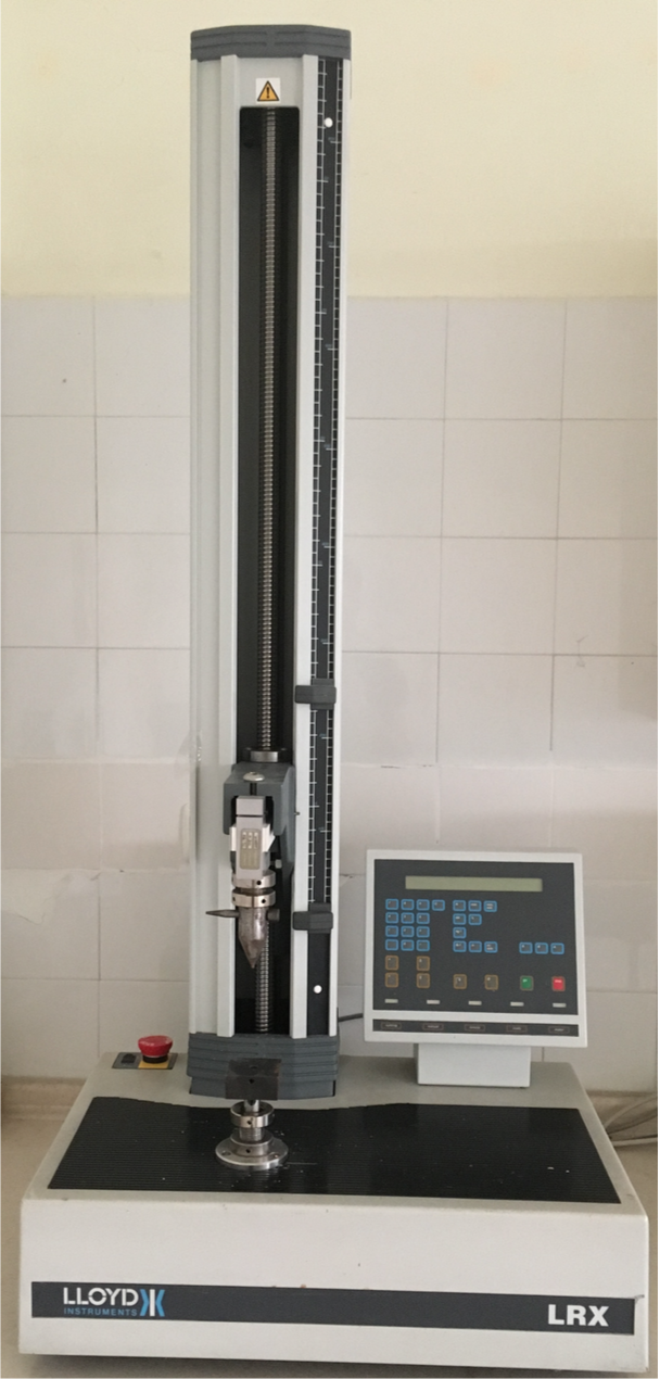
Figure 1. The push-out testing device.
Evaluation of Failure Patterns
Following the push-out test, the slices were examined under a stereomicroscope at ×40 magnification to determine the nature of bond failure. Each sample was categorized into one of the three failure modes: adhesive failure at dentin‒material interface, cohesive failure within the material, or mixed failure, which is the combination of the two failure modes (Figure 2). The operator examining the slices was blinded to which sample matched which material.

Figure 2.The images of the specimens before and after push-out test. (A) SDR group; (B) EverX Posterior group; (C) Biodentine group; (D) ProRoot MTA group; (E) Adhesive failure in SDR group; (F) Adhesive failure in EverX Posterior group; (G) Cohesive failure in Biodentine group; (H) Cohesive failure in ProRoot MTA group.
Statistical Analysis
The normality of data distribution was determined using Shapiro-Wilk test. Then the statistical differences between the groups were calculated using Kruskal-Wallis and post hoc Dunn tests. All the analyzes were performed using SPPS 21 (IBM-SPSS Inc., Chicago, IL, USA) software, and the statistical significance was set at 5%.
Results
The mean and standard deviation values of the tested materials obtained from the push-out bond strength test are presented in Table 2. SDR (4.10 ± 0.88) and EXP (3.86 ± 0.72) bulk-fill composite resins’ bond strengths were significantly higher than those of ProRoot MTA (2.29 ± 0.43) and BD (3.20 ± 0.49) calcium silicate cements (P<0.05). However, there were no significant differences between bulk-fill composite resin values and calcium silicate cement values (P>0.05).
|
Table 2. Push-out bond strength values of tested intra-orifice barrier materials (MPa)
|
|
Group
|
Mean
|
Standard Deviation
|
| ProRoot MTA |
2.29 a |
0.43 |
| Biodentine |
3.20b |
0.49 |
| SureFil SDR |
4.10b |
0.88 |
| EverX Posterior |
3.86 b |
0.72 |
|
P-value
|
< .05 |
|
a,b
Different superscript letters indicate a significant difference between groups (P<0.05).
|
The frequencies of fracture types in the test materials are presented in Table 3. No cohesive fracture was observed in SDR and EXP groups, while the mixed-type fracture was seen in the majority of cases. Mixed-type fracture was seen in 12 (40%) samples in the ProRoot MTA group and 15 (50%) samples in the BD group.
|
Table 3. Incidence of failure patterns of tested materials
|
|
Adhesive
|
Cohesive
|
Mixed
|
|
ProRoot MTA
|
16 (53.3%) |
2 (6.7%) |
12 (40%) |
|
Biodentine
|
14 (46.7%) |
1 (3.3%) |
15 (50%) |
|
SureFil SDR
|
8 (26.7%) |
0 (0%) |
22 (73.3%) |
|
EverX Posterior
|
11 (36.7%) |
0 (0%) |
19 (63.3%) |
Discussion
The cements used for endodontic purposes must tightly bind to the root canal walls and resist tooth movements or mechanic stresses that may occur during treatment procedures.20-22 For this purpose, in the present study, the push-out bond strengths of ProRoot MTA, BD, SDR and EXP materials were compared.
The bond of endodontic materials to the root dentin can be tested using different test methods such as traditional shear and push-out tests.23 Push-out test has been reported to be a reliable and practical test for examining the bond between materials and root dentin. In this test method, similar to the clinical environment, the fractures occur parallel to the resin‒dentin bond surface, and this method offers a better opportunity for analysis compared to the traditional shear test.24 For this reason, the push-out test was employed in the present study. The reason for obtaining 1-mm-thick dentin slices in the present study was that frictions that may cause misinterpretation of the results may occur in push-out test, and that it would be more reliable to utilize 1-mm-thick slices in order to minimize friction to eliminate this risk.23-25
According to the results of the present study, the push-out bond strength values of SDR, EXP and BD groups were significantly higher than those in the ProRoot MTA group. Therefore, the null hypothesis of the present study was rejected. Since there is no previous push-out bond strength study carried out using SDR and EXP bulk-fill composite resin in the literature, the results of the present study cannot be directly compared with other studies. However, the favorable outcomes of SDR might possibly be explained by its favorable stress behavior. In a recent study,17 a peculiar shrinkage behavior of the new flowable material in comparison with other flowable, nano- and micro-hybrid composites and a silorane-based material, was observed. The authors reported lower shrinkage stress, delayed point of gelation and lower shrinkage stress rates for SDR compared to the other materials that have been investigated. The high push-out bond strength of EXP material might be attributed to the ability of distribution of stresses occurring on the fibers distributed throughout the composite’s matrix.26
Similar to the present results, it has been reported in many studies that BD has significantly higher push-out bond strength values than ProRoot MTA.27-30 Moreover, Silva et al31 and Centenaro et al32 reported that BD had significantly higher push-out bond strength than MTA Angelus (Angelus, Londrina, Brazil). In another study, Alsubait et al33 examined the push-out bond strengths of white MTA (WMTA; ProRoot, Dentsply Maillefer), BD and Bio Aggregate (BA; Innovative Bio Ceramix, Vancouver, Canada) cements and reported no significant difference between MTA and BD. The reason for BD’s higher push-out bond strength compared to ProRoot MTA might be small molecular volume of BD cement and better penetration of cement into dentinal tubules, and consequently increased strength of bond to dentin. Moreover, because of this effect, the crystal formation might construct a dentin bridge within the dentinal tubules and thus the cement’s mechanic retention might increase.16,34
According to the results of present study, the fracture modes of the samples generally were mixed. The good bond of materials to dentin, because of the high push-out bond strength values exhibited by BD, SDR and EXP groups, might explain this result. Similar to the results of the present study, Centenaro et al32 reported higher incidence of mixed-type fracture after they examined the fracture modes of MTA Angelus and BD cements after push-out test. However, the incidence of adhesive failure has been reported to be higher in other studies.27,35,36 These differences in the results might be explained by differences in methodologies used in sampling and preparation.
Conclusions
Within the limitations of the present study, it might be concluded that calcium silicate-based ProRoot MTA cement’s push-out bond strength was significantly lower than those of BD, SDR and EXP materials, with no differences between the push-out bond strength values of BD, SDR and EXP.
Competing interests
The authors declare no competing interests with regards to the authorship and/or publication of this article.
Ethics approval
The study protocol was approved by Ondokuz Mayis University ethics committee.
References
- Saunders W, Saunders E. Coronal leakage as a cause of failure in root‐canal therapy: a review. Dent Traumatol 1994;10:105-8. Doi: 10.1111/j.1600-9657.1994.tb00533.x. [Crossref]
- Ray H, Trope M. Periapical status of endodontically treated teeth in relation to the technical quality of the root filling and the coronal restoration. Int Endod J 1995;28:12-8. Doi:10.1111/j.1365-2591.1995.tb00150.x. [Crossref]
- Swanson K, Madison S. An evaluation of coronal microleakage in endodontically treated teeth. Part I. Time periods. J Endod 1987;13:56-9. Doi : http://dx.doi.org/10.1016/S0099-2399(88)80135-3. [Crossref]
- Carman JE, Wallace JA. An in vitro comparison of microleakage of restorative materials in the pulp chambers of human molar teeth. J Endod 1994;20:571-5. Doi: 10.4103/1305-7456.115421. [Crossref]
- Roghanizad N, Jones JJ. Evaluation of coronal microleakage after endodontic treatment. J Endod 1996;22:471-3. Doi: 10.1016/S0099-2399(96)80080-X. [Crossref]
- Canoglu E, Gulsahi K, Sahin C, Altundasar E, Cehreli ZC. Effect of bleaching agents on sealing properties of different intraorifice barriers and root filling materials. Med Oral Patol Oral Cir Bucal 2012;17:e710. Doi: 10.4317/medoral.17751. [Crossref]
- Galvan RR, West LA, Liewehr FR, Pashley DH. Coronal microleakage of five materials used to create an intracoronal seal in endodontically treated teeth. J Endod 2002;28:59-61.
- Jacobovitz M, De Lima R. Treatment of inflammatory internal root resorption with mineral trioxide aggregate: a case report. Int Endod J 2008;41:905-912. Doi: 10.1111/j.1365-2591.2008.01412.x. [Crossref]
- Menezes R, Bramante CM, Letra A, Carvalho VGG, Garcia RB. Histologic evaluation of pulpotomies in dog using two types of mineral trioxide aggregate and regular and white Portland cements as wound dressings. Oral Surg Oral Med Oral Pathol Oral Radiol Endod 2004;98:376-9. Doi:10.1016/j.tripleo.2004.03.008. [Crossref]
- Moretton TR, Brown CE, Legan JJ, Kafrawy A. Tissue reactions after subcutaneous and intraosseous implantation of mineral trioxide aggregate and ethoxybenzoic acid cement. J Biomedic Mater Res 2000;52:528-33. Doi:5;52(3):528-33. [Crossref]
- Parirokh M, Torabinejad M. Mineral trioxide aggregate: a comprehensive literature review—part III: clinical applications, drawbacks, and mechanism of action. J Endod 2010;36:400-13. Doi: 10.1016/j.joen.2009.09.009. [Crossref]
- Dammaschke T, Gerth HU, Züchner H, Schäfer E. Chemical and physical surface and bulk material characterization of white ProRoot MTA and two Portland cements. Dent Mater 2005;21:731-8. Doi: 10.1016/j.dental.2005.01.019. [Crossref]
- Asgary S, Shahabi S, Jafarzadeh T, Amini S, Kheirieh S. The properties of a new endodontic material. J Endod 2008;34:990-3. Doi:10.1016/j.joen.2008.05.006. [Crossref]
- Gandolfi M, Pagani S, Perut F, Ciapetti G, Baldini N, Mongiorgi R, et al. Innovative silicate-based cements for endodontics: A study of osteoblast-like cell response. J Biomedic Mater Res Part A 2008;87:477-86.
- Nowicka A, Lipski M, Parafiniuk M, Sporniak-Tutak K, Lichota D, Kosierkiewicz A, et al. Response of human dental pulp capped with biodentine and mineral trioxide aggregate. J Endod 2013;39:743-7. Doi: 10.1016/j.joen.2013.01.005. [Crossref]
- Han L, Okiji T. Uptake of calcium and silicon released from calcium silicate–based endodontic materials into root canal dentine. Int Endod J 2011;44:1081-7. Doi: 10.1111/j.1365-2591.2011.01924.x. [Crossref]
- Ilie N, Hickel R. Investigations on a methacrylate-based flowable composite based on the SDR™ technology. Dent Mater 2011;27:348-55.
- Tiba A, Zeller GG, Estrich CG, Hong A. A Laboratory Evaluation of Bulk-Fill Versus Traditional Multi-Increment–Fill Resin-Based Composites. J Am Dent Assoc 2013;144:1182-3. Doi: 10.14219/jada.archive.2013.0040. [Crossref]
- Schneider SW. A comparison of canal preparations in straight and curved root canals. Oral Surg Oral Med Oral Pathol Oral Radiol 1971;32:271-5.
- Tagger M, Tagger E, Tjan AH, Bakland LK. Measurement of adhesion of endodontic sealers to dentin. J Endod 2002;28:351-4. Doi: 10.1097/00004770-200205000-00001. [Crossref]
- Huffman B, Mai S, Pinna L, Weller R, Primus C, Gutmann J, et al. Dislocation resistance of ProRoot Endo Sealer, a calcium silicate‐based root canal sealer, from radicular dentine. Int Endod J 2009;42:34-46. Doi: 10.1111/j.1365-2591.2008.01490.x. [Crossref]
- Gancedo-Caravia L, Garcia-Barbero E. Influence of humidity and setting time on the push-out strength of mineral trioxide aggregate obturations. J Endod 2006;32:894-6. Doi: 10.1016/j.joen.2006.03.004. [Crossref]
- Goracci C, Grandini S, Bossù M, Bertelli E, Ferrari M. Laboratory assessment of the retentive potential of adhesive posts: a review. J Dent 2007;35:827-35.
- Drummond J, Sakaguchi R, Racean D, Wozny J, Steinberg A. Testing mode and surface treatment effects on dentin bonding. J Biomedic Mater Res 1996;32:533-41.
- Skidmore LJ, Berzins DW, Bahcall JK. An in vitro comparison of the intraradicular dentin bond strength of Resilon and gutta-percha. J Endod 2006;32:963-6. Doi:10.1016/j.joen.2006.03.020. [Crossref]
- Garoushi S, Säilynoja E, Vallittu PK, Lassila L. Physical properties and depth of cure of a new short fiber reinforced composite. Dent Mater 2013;29:835-41. Doi: 10.1016/j.dental.2013.04.016. [Crossref]
- Guneser MB, Akbulut MB, Eldeniz AU. Effect of various endodontic irrigants on the push-out bond strength of biodentine and conventional root perforation repair materials. J Endod 2013;39:380-4. Doi: 10.1016/j.joen.2012.11.033. [Crossref]
- Nagas E, Cehreli Z, Uyanik M, Vallittu P, Lassila L. Effect of several intracanal medicaments on the push‐out bond strength of ProRoot MTA and Biodentine. Int Endod J 2016;49:184-8. Doi: 10.1111/iej.12433. [Crossref]
- EL-Ma’aita AM, Qualtrough AJ, Watts DC. The effect of smear layer on the push-out bond strength of root canal calcium silicate cements. Dent Mater 2013;29:797-803. Doi: 10.1016/j.dental.2013.04.020. [Crossref]
- Govindaraju L, Neelakantan P, Gutmann JL. Effect of root canal irrigating solutions on the compressive strength of tricalcium silicate cements. Clin Oral Investig 2016:1-5.
- Silva EJ, Carvalho NK, Zanon M, Senna PM, De-Deus G, Zuolo ML, et al. Push-out bond strength of MTA HP, a new high-plasticity calcium silicate-based cement. Braz Oral Res 2016;30. Doi: 0.1590/1807-3107BOR-2016.vol30.0084.
- Centenaro CF, Santini MF, da Rosa RA, Nascimento ALd, Kuga MC, Pereira JR, et al. Effect of calcium hydroxide on the bond strength of two bioactive cements and SEM evaluation of failure patterns. Scanning 2015.
- Alsubait SA, Hashem Q, AlHargan N, AlMohimeed K, Alkahtani A. Comparative evaluation of push-out bond strength of ProRoot MTA, bioaggregate and biodentine. J Contemp Dent Pract 2014;15:336. Doi: 1;15(3):336-40. [Crossref]
- Atmeh A, Chong E, Richard G, Festy F, Watson T. Dentin-cement Interfacial Interaction Calcium Silicates and Polyalkenoates. J Denty Res 2012;91:454-9. Doi: 10.1177/0022034512443068. [Crossref]
- Aggarwal V, Singla M, Miglani S, Kohli S. Comparative evaluation of push-out bond strength of ProRoot MTA, Biodentine, and MTA Plus in furcation perforation repair. J Conser Dent 2013;16:462. Doi: 10.4103/0972-0707.117504. [Crossref]
- Elnaghy AM. Influence of QMix irrigant on the micropush-out bond strength of biodentine and white mineral trioxide aggregate. J Adhes Dent 2014;16. Doi: 10.3290/j.jad.a320. [Crossref]