J Dent Res Dent Clin Dent Prospects. 17(4):222-226.
doi: 10.34172/joddd.2023.40483
Original Article
Assessment of peri-implant bone stress distribution with the effect of attachment type and implant location using finite element analysis
Shima Aalaei Conceptualization, Methodology, Project administration, Writing – original draft, Writing – review & editing, 1 
Atefeh Sheikhi Data curation, Writing – original draft, Writing – review & editing, 2
Parisa Mehdian Formal analysis, Investigation, Visualization, Writing – original draft, Writing – review & editing, 2
Farnoosh Taghavi Resources, Writing – original draft, Writing – review & editing, 3
Sara Salimian Data curation, Writing – original draft, Writing – review & editing, 4
Farnaz Taghavi-Damghani Supervision, Validation, Writing – original draft, Writing – review & editing, 5, 3, * 
Author information:
1Department of Prosthodontics, Dental Caries Prevention Research Center, Qazvin University of Medical Sciences, Qazvin, Iran
2Student Research Committee, Qazvin University of Medical Sciences, Qazvin, Iran
3Department of Prosthodontics, School of Dentistry, Shahid Beheshti University of Medical Sciences, Tehran, Iran
4Student Research Committee, Tehran University of Medical Sciences, Tehran, Iran
5Dental Caries Prevention Research Center, Qazvin University of Medical Sciences, Qazvin, Iran
Abstract
Background.
The objective of the current research was to evaluate how stress is distributed in the peri-implant bone of a mandibular overdenture with implants placed asymmetrically to the midline.
Methods.
A 26-year-old male’s mandible, with missing teeth, was examined using computed tomography (CT) scanning. Two implants were inserted at right angles to the occlusal plane, in the positions of the right canine and left lateral incisor of the mandible, with an internal connection. Two types of attachments (bar and ball) were designed. To simulate the clinical condition, anterior (on central incisors) and bilateral posterior (on premolars and molars) loadings were applied. The stress distribution was assessed using finite element analysis (FEA).
Results.
The lateral incisor level implant was found to have the highest maximum principal stress (about 33 MPa) in both models in the anterior loading condition. However, in both models, the canine-level implant revealed more stress values (about 22 MPa) in the posterior loading condition.
Conclusion.
In mandibular implant-supported overdentures, when implants were placed asymmetrically to the midline, one acted as a fulcrum and sustained more occlusal load. The bar attachment system did not reveal superior results in terms of stress distribution compared to the ball attachment.
Keywords: Dental implants, Finite element analysis, Overdenture, Stress
Copyright and License Information
©2023 The Author(s).
This is an open-access article distributed under the terms of the Creative Commons Attribution License (
http://creativecommons.org/licenses/by/4.0), which permits unrestricted use, distribution, and reproduction in any medium provided the original work is properly cited.
Introduction
Implant-supported overdentures have been introduced to provide esthetics and functional rehabilitation for patients wearing complete dentures.1,2 The high success rate of dental implants has led to the selection of an overdenture based on two implants as one of the treatment options in the edentulous mandible.3
There are splinted (bar attachments) and non-splinted (ball, locator, etc.) anchorage systems that can improve the retention and stability of an implant-supported overdenture.4 Attachment selection is one of the challenges among clinicians and depends on multiple factors such as retention, occlusal space, and jaw anatomy.5
Attachments transfer the stress from mastication to the implants, and clinicians should consider the stress values to be in a safe range.6 Ball attachments are simple to use and cost-effective and can reduce the occlusal force by absorbing the loading stress. Patients can clean this type of attachment easily.5,7 More vertical restorative space is needed for the bar and clip attachment compared to the non-splinted ones.5 The stress values can be reduced using the bar attachment, as suggested by Misch,8 due to the splinting ability.
Finite element analysis (FEA) is one of the methods to assess stress distribution in bone‒implant systems. It has several advantages, such as generating complex models and analyzing internal stress accurately.9,10
The failure of an implant system is greatly influenced by the level of stress concentration in the peri-implant bone.6 Misch8 suggested that the loading forces would be reduced by placing the implants at the same occlusal height and symmetrically from the midline. A few studies have evaluated the stress distribution in models with different implant positions.11,12 Alvarez-Arenal et al3 reported that implants at the premolar level had better stress distribution than implants at the lateral incisor and canine levels. No evidence was found to compare asymmetrical implants from midline with different types of attachments. Hence, the innovation of this study is that the effect of two parameters with different loading conditions was analyzed. This study assessed and compared stress distribution in the bone adjacent to the implants placed asymmetrically from the midline with ball and bar attachments.
Methods
A study model was created using a computed tomography (CT) scan of a 26-year-old man’s edentulous mandible to evaluate how stress is distributed in the bone around the dental implants.
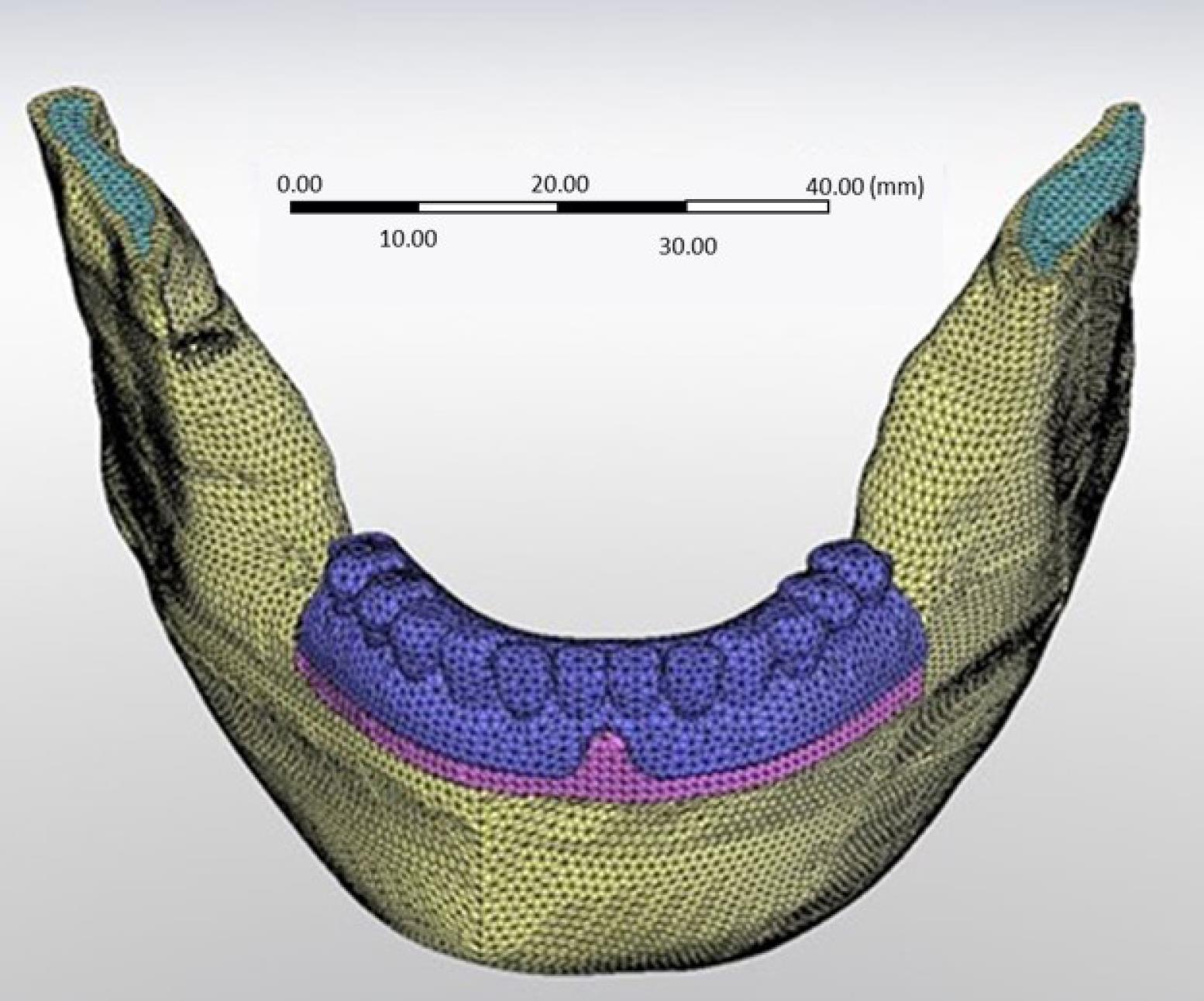
Data were processed with an image processing software, MIMICS (Materialise Interactive Medical Image Control System; Materialise, version 21, Leuven, Belgium). Then, the image was transferred to SolidWorks software (version 28, Dassault Systems SolidWorks Corp., MA, United States). Two bone-level implants (ITI, Straumann, Switzerland, 10 × 4.1 mm) were modeled and placed in the right canine and left lateral incisor region. The Dolder bar (3.25 mm height) and ball attachments (3.4 mm height) were inserted in two separate models (Figure 1). The bone thickness was extracted from the CT scans, and a mucosal membrane with a 2-mm thickness was modeled using SolidWorks software. All the simulated materials were considered homogeneous with a linear modulus of elasticity. The final model was meshed, taking into account the physical properties of the materials (Table 1) and the boundary conditions (Figure 2). The ball model had 468 472 elements, while the bar model had 150 566 elements.

Figure 1.
3D models of the A) bar and B) ball attachments
.
3D models of the A) bar and B) ball attachments
Table 1.
Physical properties of the materials
|
Materials
|
Elastic modulus (MPa)
|
Poisson ratio
|
Reference
|
| Cortical bone |
13700 |
0.3 |
3,13
|
| Acrylic resin |
3000 |
0.35 |
14
|
| Trabecular bone |
1370 |
0.3 |
3
|
| Mucosa |
680 |
0.45 |
14
|
| Ball abutment and metallic cap |
114000 |
0.3 |
3
|
| Implant |
110000 |
0.33 |
15
|
| Bar |
218000 |
0.33 |
16,17
|
| Clips |
3000 |
0.28 |
18
|
| Lamella retention insert |
97000 |
0.42 |
6
|

Figure 2.
Meshing process
.
Meshing process
The total number of nodes in the bar model was 223 173, and in the ball model was 746 557. The size of each element was 1.5 mm. Parabolic and tetrahedral solid elements were used.
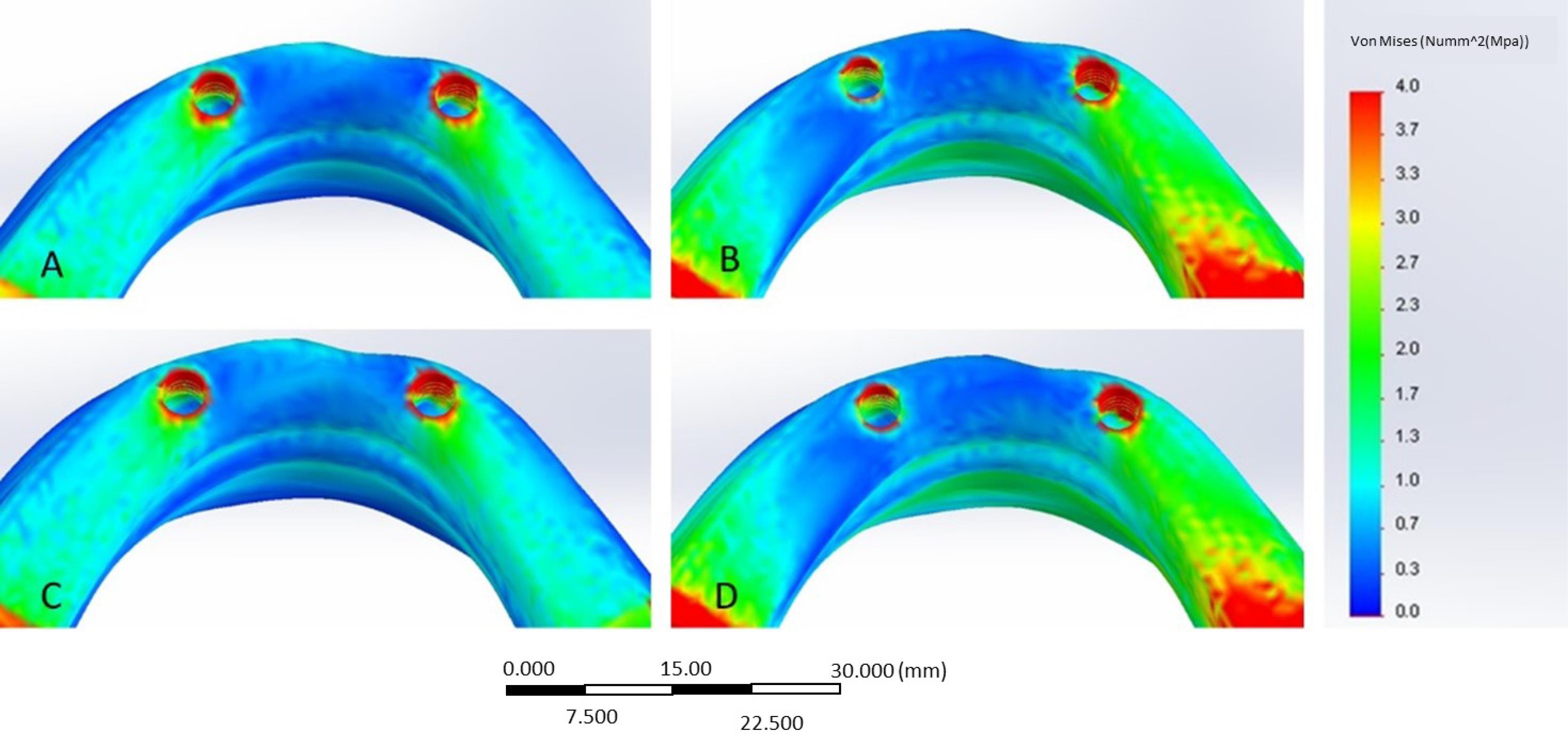
The COSMOWorks software (version 12.1., Dassault Systems SolidWorks Corp., MA, United States) was used to model the masseter and internal pterygoid muscle and apply a loading condition on the anterior (central and lateral incisors) and posterior (molars and premolars) regions (Figure 3). The amount of muscle force in each condition was based on the studies of Korioth and Hannam13 and determined by the multiplication of two parameters, namely weight factor and scaling factor (Table 2). The stress values were analyzed and described in color-coded figures (Figure 4).

Figure 3.
Modeling of the muscles
.
Modeling of the muscles
Table 2.
The forces of the muscles (weight factor and scaling factor)
|
|
Weight factor (Newton)
|
Scaling factor
|
|
Anterior clenching
|
Posterior clenching
|
|
Right
|
Left
|
Right
|
Left
|
| Superficial masseter |
190.4 |
0.40 |
0.40 |
1.00 |
1.00 |
| Deep masseter |
81.6 |
0.26 |
0.26 |
1.00 |
1.00 |
| Medial pterygoid |
174.8 |
0.78 |
0.78 |
0.76 |
0.76 |

Figure 4.
Stress values in the A) bar model - anterior loading, B) bar model - bilateral posterior loading, C) ball model - anterior loading, and D) ball model – bilateral posterior loading
.
Stress values in the A) bar model - anterior loading, B) bar model - bilateral posterior loading, C) ball model - anterior loading, and D) ball model – bilateral posterior loading
Results
In the present study, the stress distribution in the peri-implant bone was measured using FEA in different loading conditions (Table 3). In the bar model, by applying anterior loads, the cortical bone of the lateral incisor-level implant exhibited the maximum stress (33.3 MPa). The canine-level implant showed a stress value of 22.7 MPa in this loading condition. The canine-level implant had the highest maximum principal stress (22 MPa) by bilateral posterior loading application. The cortical bone surrounding the lateral incisor-level implant had a stress value of 20.7 MPa.
Table 3.
Maximum stress values (MPa) with bar and ball attachments
|
|
|
Anterior loading
|
Posterior bilateral loading
|
| Bar attachment |
Right implant (canine) |
22.7 |
22 |
| Left implant (lateral incisor) |
33.3 |
20.7 |
| Ball attachment |
Right implant |
27.4 |
22.2 |
| Left implant |
33.2 |
17.9 |
The cortical bone of the implant at the level of the lateral incisor had the highest principal stress (33.2 MPa) when anterior loads were applied in the ball model. The adjacent bone stress value at the canine-level implant measured 27.4 MPa.
The stress value in the bone adjacent to the canine-level implant was 27.4 MPA. Under bilateral posterior clenching, the maximum principal stress was 22.2 MPa in the canine-level peri-implant bone, and 17.9 MPa was recorded adjacent to the lateral incisor-level implant. Therefore, the distal implant exhibited the highest stress concentration in both models when subjected to posterior loads, and with anterior loads, the medial implant in both models exhibited more stress.
Discussion
In this study, the stress was analyzed in the bone adjacent to the asymmetrically placed implants. No similar study was found in the literature to compare the results. Studies in biomechanics have demonstrated that the primary cause of crestal bone loss and implant failure shortly after the implant is loaded is primarily due to a great deal of stress at the implant‒bone interface.18,19 In the present study, the greatest stress concentration was found in the cortical bone surrounding the implant neck due to the higher density and modulus of elasticity of cortical bone compared to that of trabecular bone.
The findings regarding the use of splinted or non-splinted attachments have controversies in different articles.8,20,21 Misch8 suggested that the bar attachment can distribute the stress more evenly compared to the ball attachment. Satpathy et al20 showed that ball attachment could be a favorable system in conditions with a low range of force. However, a bar/clip attachment may have better results when a higher force range is expected. Park et al21 assessed the effect of attachment type and palatal coverage in the maxillary implant-supported overdentures. They concluded that ball attachment revealed better stress distribution than that of the attachment of the milled bar.
In this study, both models showed similar stress levels in the peri-implant bone adjacent to the area where the load is applied. However, in the peri-implant bone far from the place of load application with anterior loads, the model with the bar attachment showed less stress. With posterior loads, the model with ball attachments exhibited lower stress values due to the rotational movements around the ball attachments.
According to the present study, the bar attachment system did not reveal superior results in stress distribution compared to the ball attachment in asymmetrically placed implants. The ball attachment allows a wide motion range for the prosthesis and absorbs stress. This free-rotating motion of overdenture increases the force distribution in the mucosal tissue and reduces the stress accumulation in the implant and the surrounding bone.22 Bar attachment splint fixtures increase the retention and reduce the range of motion.8,23 Therefore, the prosthesis has limited anteroposterior movements with bilateral posterior loads. In this study, it can be discussed that with anterior loads, the labial part of the anterior alveolar bone in both models prevented the continued rotation of the prosthesis. Hence, the effect of bar attachment in terms of stress distribution was greater than the ball attachment.
Misch8 described that implants should be placed symmetrically from the midline. When one implant is farther from the midline, it will act as the rotation point during posterior load application. However, the anterior implant will act as a fulcrum and show higher stress in the anterior bite condition, consistent with the present study.
FEA is a numerical method with great efficiency in stress distribution analysis. This method has the advantage of simulating models with complex geometries and the possibility of changing mechanical parameters. Modeling of trabecular and cortical bone to assess the stress patterns is difficult due to the heterogeneous structure and several factors such as age, gender, and type of the bone.12,23,24 In this study, the mechanical properties of the bone were assumed to be isotropic and homogeneous, based on the methods used in several previous research.12,24
The data obtained from this study provide information on the precise areas where stress is concentrated. However, it is necessary to conduct long-term clinical studies to determine the exact stress values and the effect of loadings on the surrounding tissues.
Conclusion
According to the results, better stress distribution was not achieved by using splinted attachments compared to the non-splinted attachments. The model with the bar attachment showed less stress in the peri-implant bone far from the place of load application with anterior loads, and the model with ball attachments exhibited lower stress values due to the rotational movements around the ball attachments with posterior loads. When implants were placed asymmetrically in the mandible, one implant acted as a fulcrum after applying occlusal loads. The distal implant exhibited more stress values under posterior loading; the medial implant showed greater stress values under anterior loads.
Competing Interests
The authors declare that they have no conflicts of interest.
Data Availability Statement
The data used to support the findings of this study are included in the article.
Ethical Approval
This study was approved by the Ethics Committee of Qazvin University of Medical Sciences with an ethical number of IR.QUMS.REC.1394.682. There was no conflict with ethical considerations.
Funding
This research received no specific grant from funding agencies in the public, commercial, or not-for-profit sectors.
References
- Unsal GS, Erbasar GNH, Aykent F, Ozyilmaz OY, Ozdogan MS. Evaluation of stress distribution on mandibular implant-supported overdentures with different bone heights and attachment types: a 3D finite element analysis. J Oral Implantol 2019; 45(5):363-70. doi: 10.1563/aaid-joi-D-19-00076 [Crossref] [ Google Scholar]
- Patil PG, Seow LL, Uddanwadikar R, Ukey PD. Biomechanical behavior of mandibular overdenture retained by two standard implants or 2 mini implants: a 3-dimensional finite element analysis. J Prosthet Dent 2021;125(1):138.e1-138.e8. 10.1016/j.prosdent.2020.09.015.
- Alvarez-Arenal A, Gonzalez-Gonzalez I, deLlanos-Lanchares H, Brizuela-Velasco A, Martin-Fernandez E, Ellacuria-Echebarria J. Influence of implant positions and occlusal forces on peri-implant bone stress in mandibular two-implant overdentures: a 3-dimensional finite element analysis. J Oral Implantol 2017; 43(6):419-28. doi: 10.1563/aaid-joi-D-17-00170 [Crossref] [ Google Scholar]
- El-Anwar MI, El-Taftazany EA, Hamed HA, ElHay MAA. Influence of number of implants and attachment type on stress distribution in mandibular implant-retained overdentures: finite element analysis. Open Access Maced J Med Sci 2017; 5(2):244-9. doi: 10.3889/oamjms.2017.047 [Crossref] [ Google Scholar]
- Ebadian B, Talebi S, Khodaeian N, Farzin M. Stress analysis of mandibular implant-retained overdenture with independent attachment system: effect of restoration space and attachment height. Gen Dent 2015; 63(1):61-7. [ Google Scholar]
- Shishesaz M, Ahmadzadeh A, Baharan A. Finite element study of three different treatment designs of a mandibular three implant-retained overdenture. Lat Am J Solids Struct 2016; 13(16):3126-44. doi: 10.1590/1679-78253212 [Crossref] [ Google Scholar]
- Khurana N, Rodrigues S, Shenoy S, Saldanha S, Pai U, Shetty T. A comparative evaluation of stress distribution with two attachment systems of varying heights in a mandibular implant-supported overdenture: a three-dimensional finite element analysis. J Prosthodont 2019; 28(2):e795-e805. doi: 10.1111/jopr.12966 [Crossref] [ Google Scholar]
- Misch CE. Dental Implant Prosthetics. Philadelphia, PA: Elsevier; 2015.
- Amaral CF, Gomes RS, Rodrigues Garcia RCM, Del Bel Cury AA. Stress distribution of single-implant-retained overdenture reinforced with a framework: a finite element analysis study. J Prosthet Dent 2018; 119(5):791-6. doi: 10.1016/j.prosdent.2017.07.016 [Crossref] [ Google Scholar]
- Mozaffari A, Hashtbaran D, Moghadam A, Aalaei S. Stress distribution in peri-implant bone in the replacement of molars with one or two implants: a finite element analysis. J Dent (Shiraz) 2023; 24(1 Suppl):132-7. doi: 10.30476/dentjods.2022.92584.1659 [Crossref] [ Google Scholar]
- Alvarez-Arenal A, Gonzalez-Gonzalez I, deLlanos-Lanchares H, Martin-Fernandez E, Brizuela-Velasco A, Ellacuria-Echebarria J. Effect of implant- and occlusal load location on stress distribution in Locator attachments of mandibular overdenture A finite element study. J Adv Prosthodont 2017; 9(5):371-80. doi: 10.4047/jap.2017.9.5.371 [Crossref] [ Google Scholar]
- Aalaei S, Abedi P, Niknami S, Taghavi F, Taghavi-Damghani F. The effect of attachment types and implant level on the stress distribution in a mandibular overdenture: a 3D finite element analysis. Braz Dent Sci 2022; 25(3):e3355. doi: 10.4322/bds.2022.e3355 [Crossref] [ Google Scholar]
- Korioth TW, Hannam AG. Deformation of the human mandible during simulated tooth clenching. J Dent Res 1994; 73(1):56-66. doi: 10.1177/00220345940730010801 [Crossref] [ Google Scholar]
- Ozan O, Ramoglu S. Effect of implant height differences on different attachment types and peri-implant bone in mandibular two-implant overdentures: 3D finite element study. J Oral Implantol 2015; 41(3):e50-9. doi: 10.1563/aaid-joi-d-13-00239 [Crossref] [ Google Scholar]
- Shahmiri R, Das R. Finite element analysis of implant-assisted removable partial denture attachment with different matrix designs during bilateral loading. Int J Oral Maxillofac Implants 2016; 31(5):e116-27. doi: 10.11607/jomi.4400 [Crossref] [ Google Scholar]
- Barão VA, Assunção WG, Tabata LF, Delben JA, Gomes EA, de Sousa EA. Finite element analysis to compare complete denture and implant-retained overdentures with different attachment systems. J Craniofac Surg 2009; 20(4):1066-71. doi: 10.1097/SCS.0b013e3181abb395 [Crossref] [ Google Scholar]
- Hussein MO. Stress-strain distribution at bone-implant interface of two splinted overdenture systems using 3D finite element analysis. J Adv Prosthodont 2013; 5(3):333-40. doi: 10.4047/jap.2013.5.3.333 [Crossref] [ Google Scholar]
- Assunção WG, Tabata LF, Barão VA, Rocha EP. Comparison of stress distribution between complete denture and implant-retained overdenture-2D FEA. J Oral Rehabil 2008; 35(10):766-74. doi: 10.1111/j.1365-2842.2008.01851.x [Crossref] [ Google Scholar]
- Joshi S, Kumar S, Jain S, Aggarwal R, Choudhary S, Reddy NK. 3D finite element analysis to assess the stress distribution pattern in mandibular implant-supported overdenture with different bar heights. J Contemp Dent Pract 2019; 20(7):794-800. [ Google Scholar]
- Satpathy S, Babu CL, Shetty S, Raj B. Stress distribution patterns of implant supported overdentures-analog versus finite element analysis: a comparative in-vitro study. J Indian Prosthodont Soc 2015; 15(3):250-6. doi: 10.4103/0972-4052.165324 [Crossref] [ Google Scholar]
- Park JH, Wang YK, Lee JJ, Park YH, Seo JM, Kim KA. Effect of attachments and palatal coverage of maxillary implant overdenture on stress distribution: a finite element analysis. J Dent Rehabil Appl Sci 2020; 36(2):70-9. doi: 10.14368/jdras.2020.36.2.70 [Crossref] [ Google Scholar]
- Metwally AF, Gamal M. Evaluation of stresses distribution pattern in implant- retained maxillary obturators using nova-lock, ball & locator-bar attachment systems (Three dimensional finite element analysis). Egypt Dent J 2019; 65(3):2597-605. doi: 10.21608/edj.2019.72623 [Crossref] [ Google Scholar]
- Anas El-Wegoud M, Fayyad A, Kaddah A, Nabhan A. Bar versus ball attachments for implant-supported overdentures in complete edentulism: a systematic review. Clin Implant Dent Relat Res 2018; 20(2):243-50. doi: 10.1111/cid.12551 [Crossref] [ Google Scholar]
- Taghavi-Damghani F, Salari AM, Shayegh SS, Taghavi F. Effect of abutment height difference on stress distribution in mandibular overdentures: a three-dimensional finite element analysis. J Iran Dent Assoc 2022; 34(1-2):1-8. [ Google Scholar]