J Dent Res Dent Clin Dent Prospects. 18(2):129-134.
doi: 10.34172/joddd.40900
Original Article
Effect of natural silk fibers and synthetic fiber-reinforced composites on the cytotoxicity of fibroblast cell lines
Mutiara Annisa Conceptualization, Data curation, Investigation, Project administration, Writing – original draft, Writing – review & editing, 
Dyah Irnawati Data curation, Formal analysis, Investigation, Methodology, Validation, Writing – original draft, Writing – review & editing, 
Widowati Siswomihardjo Formal analysis, Investigation, Methodology, Validation, Writing – original draft, Writing – review & editing, 
Siti Sunarintyas Conceptualization, Data curation, Formal analysis, Investigation, Project administration, Supervision, Writing – original draft, Writing – review & editing, * 
Author information:
Department of Dental Biomaterials, Faculty of Dentistry, Universitas Gadjah Mada, Yogyakarta,Indonesia
Abstract
Background.
Synthetic fibers have many benefits in clinical practice; however, they cause microplastic pollution, and their unaffordable price increases treatment costs. Natural silk fibers require biocompatibility assessment. This study investigated the effects of natural and synthetic fiber-reinforced composites (FRCs) on the cytotoxicity of fibroblast cell lines.
Methods.
Three commercial synthetic fibers (polyethylene, quartz, and E-glass) and two silk fibers from Bombyx mori and Samia ricini cocoons were employed. These fibers were made into FRC samples (n=6) by impregnation in flowable composite using a brass mold (25×2×2 mm). NIH/3T3 mouse fibroblasts were cultured in Dulbecco’s modified eagle medium, supplemented, and seeded in 2×10⁴ cells/mL. They were stored at 37 °C under 5% CO2 for 24 hours. The FRC samples were made into powder, eluted in dimethylsulfoxide, continued with PBS, supplemented with Dulbecco’s modified eagle medium (DMEM), and exposed to cells for 24 hours. Blank (medium only) and control (cells and medium) samples were included. Subsequently, MTT was added for 4 h and read by enzyme-linked immunosorbent assay (λ=570 nm). Cell viability (%) was calculated and analyzed using one-way ANOVA (α=0.05).
Results.
All groups of FRCs showed>80% cell viability. One-way ANOVA showed no significant difference between FRC groups regarding the viability of fibroblast cell lines (P>0.05).
Conclusion.
Both natural silk and synthetic fibers exhibit low cytotoxicity to fibroblast cell lines. B. mori and S. ricini silk fibers showed the potential to be used as alternative synthetic fibers.
Keywords: Bombyx mori, Cytotoxicity, Fiber-reinforced composites, Fibroblast cell lines, Samia ricini
Copyright and License Information
©2024 The Author(s).
This is an open access article distributed under the terms of the Creative Commons Attribution License (
http://creativecommons.org/licenses/by/4.0/), which permits unrestricted use, distribution, and reproduction in any medium, provided the original work is properly cited.
Introduction
Fibers have been widely used in dental clinical settings as fiber-reinforced composites (FRCs), mostly for applications such as dental restorations, periodontal splints, endodontic posts, orthodontic retainers, fixed partial dentures, and removable dentures.1 The use of FRCs relies on their exceptional properties, such as physicomechanical, bonding, and viscoelastic capabilities and biocompatibility, which are set forth by the specific fiber and matrix used.1,2 In addition, using FRCs in clinical practice enables minimal preparation of the neighboring tooth structure.2
Dental composites are strengthened using various fibers, including synthetic materials such as polyethylene, carbon, glass, Kevlar (p-phenylene diamine), and plant- and animal-derived natural fibers. These fibers can be arranged in unidirectional, braided, and woven patterns.1,3 Although synthetic fibers offer benefits such as durability, high tensile strength, elastic modulus, and ultimate strain in FRCs, their use is often expensive, leading to high dental treatment costs.4,5 Furthermore, this synthetic material is nonbiodegradable, is not eco-friendly, and contributes to microplastic contamination. To address these limitations, interest is growing in natural fibers as substitutes for synthetic fibers. These natural fibers can be cellulose-rich plant or animal fibers mainly made up of proteins.6 Silk fiber is considered a potential animal-derived fiber because its mechanical qualities are superior to those of plant fibers, and its specific mechanical performance is comparable to those of glass and polyethylene fibers.5,7 The silk fiber that can be used originated from Bombyx mori, a controlled silkworm fed with mulberry. In a study by Shah et al,8 the tensile strength and specific strength of a composite laminate made from B. mori silk fiber and glass fiber were comparable. This finding is consistent with those reported by Sunarintyas et al,5 who used an identical silk fiber material and demonstrated a flexural strength similar to that of polyethylene FRCs. Despite limited evidence, the potential use of the non-mulberry cocoon type is exemplified by Attacus atlas, Cricula trifenestrata, and Samia ricini in terms of the quality of the silk fiber and its protein components, such as sericin and fibroin.7,9,10
However, the biocompatibility issue regarding the effect of FRC use on biological tissue is widely discussed because FRCs have the same resin matrix as conventional resin composites, such as methyl methacrylate (MMA), which may cause allergy, contact dermatitis, and mucous membrane irritation.11,12 Incorporating fiber and resin matrix also contributes to chemical reactions that affect cytocompatibility.6,10 ISO 10993-1 suggests appropriate steps for the preliminary assessment of biological compatibility of medical devices through in vitro evaluation of cytotoxicity.10,13 Thus, this study aimed to investigate the effects of natural and synthetic FRCs on the cytotoxicity of fibroblast cell lines.
Methods
This experimental laboratory study received ethical clearance from the Ethics and Advocacy Commission of the Faculty of Dentistry at Universitas Gadjah Mada (Approval No. 73/UN1/KEP/FKG-RSGM/EC/2023). The NIH/3T3 mouse fibroblast cell line (ATCC, Old Town, MD, USA) was used in the cytotoxicity assay. Cell culture was performed using Dulbecco’s modified eagle medium (DMEM; Invitrogen, Carlsbad, CA, USA). The supplementation included fetal bovine serum (FBS, Sigma-Aldrich, USA), penicillin (Gibco, Grand Island, NY, USA), and gentamicin (Gibco). Five types of fibers consisting of two types of natural silk fibers and three types of synthetic fibers were employed. The details of the materials used for specimen preparation are provided in Table 1.
Table 1.
Information on the materials used for specimen making in this study
|
Materials
|
Manufacturer
|
Description
|
Number of specimens
|
|
Bombyx mori silk fiber |
Local silk from Wajo, South Sulawesi, Indonesia |
Domesticated silk fiber derived from the cocoon of Bombyx mori, tailored into silk ribbon in unidirectional configuration |
6 |
|
Samia ricinisilk fiber |
Local silk from Kulon Progo, Yogyakarta, Indonesia |
Wild silk fiber derived from the cocoon of Samia ricini, tailored into silk ribbon in unidirectional configuration |
6 |
| Polyethylene fiber |
Kerr Construct, USA |
Commercially available synthetic fiber made of UHMWPE in woven configuration |
6 |
| Quartz fiber |
Quartz Splint, France |
Commercially available synthetic fiber made of quartz in unidirectional configuration |
6 |
| E-glass fiber |
everStickTM, Stick Tech Ltd, Finland |
Commercially available synthetic fiber made of E-glass impregnated with bis-GMA and PMMA in unidirectional configuration |
6 |
| Flowable composite resin |
DenFil Flow, Vericom, Korea |
Light-cured flowable composite resin used to make specimens of FRCs |
- |
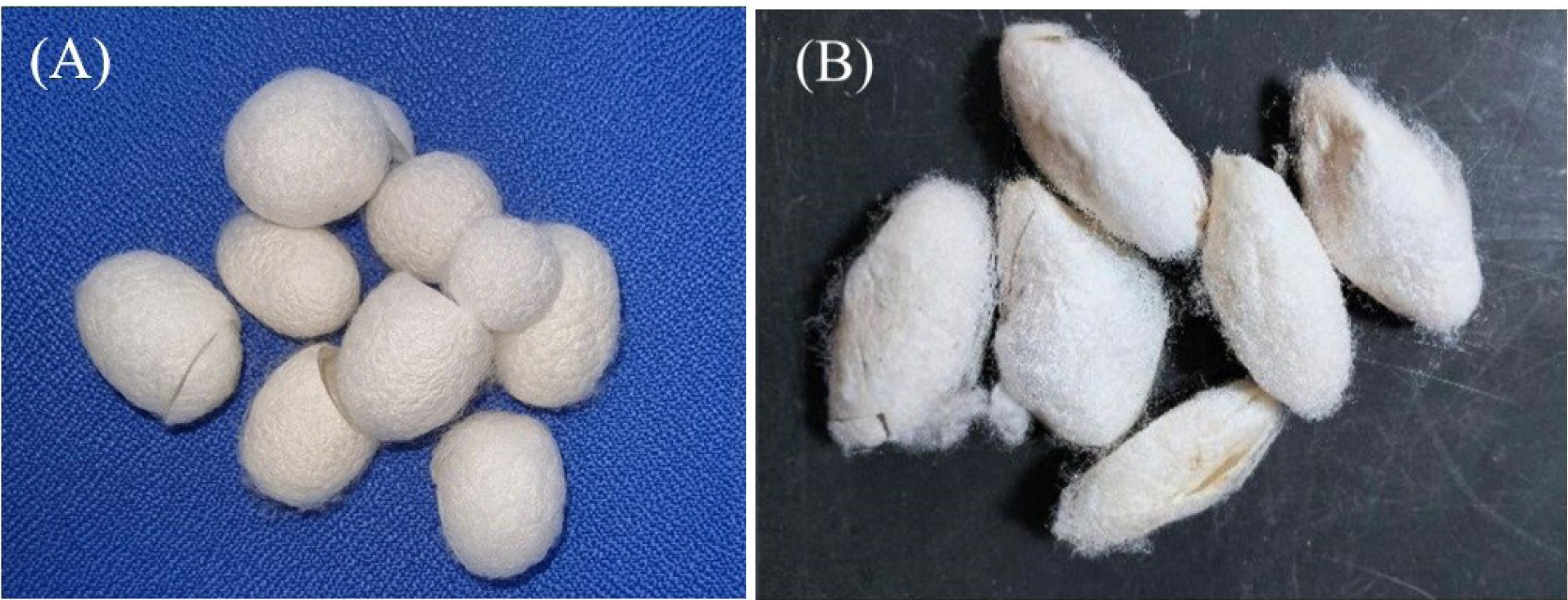
Table 1 presents the specifications of the natural and synthetic fibers used in this study. While synthetic fibers are readily available, both natural silk fibers must be tailored before they can be transformed into FRC specimens. Initially, the cocoons of B. mori and S. ricini (Figure 1) were subjected to a degumming technique, as described by Rameshbabu et al,14 to remove sericin. The silk fibers were then extracted and twisted into threads.

Figure 1.
Cocoon of silkworm. (A) Bombyx mori and(B) Samia ricini
.
Cocoon of silkworm. (A) Bombyx mori and(B) Samia ricini
Preparation of the silk ribbon
The unidirectional silk ribbons from each silk fiber were prepared following the protocol of Sunarintyas et al.5 Silk fibers of B. mori and S. riciniwere weighed to 0.155 g each and then arranged in a brass mold (80 × 2 mm), followed by impregnation with flowable composite resin. The resulting silk ribbon was stored at 4 °C.
Specimen preparation
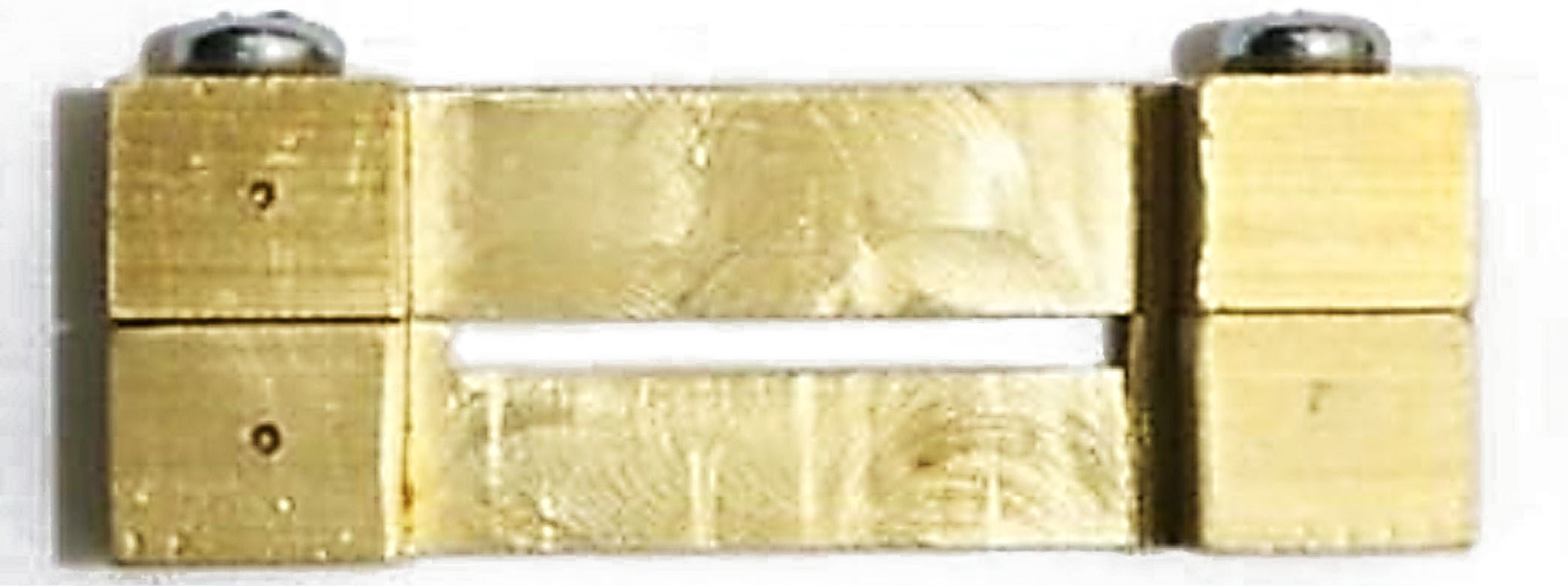
The specimens were prepared according to the procedure outlined by Frese et al,15 with a few modifications. Aseptically, a brass mold measuring 25 × 2 × 2 mm was positioned and secured onto a microscope slide. A layer of flowable composite resin was applied to the lower portion of the mold, which occupied roughly one-third of its height. Subsequently, each fiber was inserted into the mold and saturated with the composite resin. After that, another layer of composite resin was added to fill the mold. Subsequently, they were delicately compressed using a different microscope slide and subjected to light curing following the manufacturer’s guidelines. Figure 2 is a schematic representation of the sample preparation process.

Figure 2.
The sample preparation process
.
The sample preparation process
Cell culture
Cell culture was performed according to the method employed by Klein-Junior et al.16 NIH/3T3 murine fibroblasts were regularly cultured in DMEM. The solution was enriched with 10% fetal bovine serum, 100 U/mL penicillin, 100 U/mL streptomycin, and 100 μg/mL gentamicin. The samples were arranged in 96-well plates, and the cells were distributed onto the samples at a concentration of 2 × 10⁴ cells/mL. The cells were subsequently placed in a humidified incubator at 37°C with a carbon dioxide concentration of 5% for 24 hours.
Preparation of the stock solution
Before the cytotoxicity test, all FRC specimens were stored in a sterile saline solution for 24 hours at room temperature. Each group of FRCs had six specimens (n = 6). Subsequently, the fibers were pulverized and dissolved in dimethylsulfoxide, further diluted with PBS, and supplemented with DMEM to achieve a stock solution concentration of 100 µg/mL for each fiber type. The stock solutions were filtered using a 0.22-µm Millipore membrane (Millipore; Billerica, MA, USA) and stored at 4 °C.12
Cell viability
The 3-(4,5-dimethylthiazol-2-yl) 2,5-diphenyl tetrazolium bromide (MTT) cytotoxicity test was performed according to ISO 10993-1. A fibroblast cell line consisting of 2 × 10⁴ cells/mL in supplemented DMEM was placed in 96-well plates and cultured for 24 hours until a semiconfluent monolayer was formed. Subsequently, they were subjected to several fiber stock solutions, with six replications for each group (n = 6). The medium without cells was used as the blank, whereas the medium with cells was utilized as the control. The incubation period for the treated and untreated groups lasted 24 hours at 37 °C with a CO2 concentration of 5%. Following a 24-hour exposure period, the MTT assay was performed by introducing MTT and allowing it to incubate. Following a 4-hour incubation period, formazan formation was assessed for each treatment concentration using an enzyme-linked immunosorbent assay reader set at 570 nm. The cell viability percentage in the treated cells was determined relative to that in the control cells using the following formula:
Statistical analysis
Data were reported as the average percentage of cell viability ± standard deviation. The effect of different types of FRCs on the viability of fibroblasts was determined through statistical analysis using ANOVA.
Results
The cell viability of the fibroblast cell lines in the treated group was compared with that of the control cells (untreated group) (Table 2).
Table 2.
Cell viability percentage of NIH/3T3 fibroblast cell line after 24 hours of exposure to various fiber-reinforced composites (FRCs)
|
Groups
|
Cytoviability (%)±SD
|
| Fibroblast cell lines (control) |
100.00 ± 35.47 |
|
Bombyx mori silk FRCs |
100.44 ± 16.35 |
|
Samia ricinisilk FRCs |
114.47 ± 27.70 |
| Polyethylene FRCs |
103.07 ± 28.77 |
| Quartz FRCs |
102.41 ± 38.29 |
| E-glass FRCs |
118.20 ± 38.29 |
Table 2 indicates that all the categories of FRCs had cell viability percentages exceeding 80%, surpassing the percentage of the control group, which included only the medium and cells. Statistical analysis was extended using one-way ANOVA. The results (Table 3) indicated the lack of significant variations in the viability of the fibroblast cell line after 24 hours of exposure to all FRCs (P > 0.05).
Table 3.
ANOVA summary of the cell viability of NIH/3T3 fibroblast cell line after 24 hours of exposure to various FRCs
|
|
Sum of square
|
df
|
Mean square
|
F
|
P
value
|
| Between groups |
1381.980 |
4 |
345.495 |
0.401 |
0.806 |
| Within groups |
21529.037 |
25 |
861.161 |
|
|
| Total |
22911.017 |
29 |
|
|
|
Discussion
Composites are artificial materials with multiple phases and a desirable combination of the most advantageous features from each phase.17 When polymers are used to reinforce composites, resulting in polymer matrix composites, they can be classified into two types: particle-reinforced types, such as dental composites, and fiber-reinforced types, such as dental FRCs. Fibers can be composed of various materials, including carbon, aramid, polyethylene, or glass, which fall into the categories of synthetic fibers. In addition, natural fibers are derived from plants, such as jute, coir, and sisal, and animal fibers, such as wool and silk.18 This material is designed to possess exceptional qualities for various therapeutic uses. However, potential changes and reactions that may occur in terms of biocompatibility must be considered.19
This study examined the cytotoxic effects of synthetic and natural FRCs on a fibroblast cell line. Multiple in vitro test paradigms are available for assessing the cytotoxicity of dental biomaterials. The methods used include direct contact, where the materials come into direct contact with the cellular layer; indirect contact, where a barrier is placed between the cell and the biomaterial layer, such as agar overlay assay and filter diffusion; and extract method, where the extracts of materials are placed in contact with cells.13 An ideal in vitro test closely replicates the in vivo settings. However, certain factors must be considered, such as the nature of the biomaterial being examined (substances, solid, and powder) and the specific purpose of the test being undertaken. This study used the direct contact assay, which is the most sensitive method for assessing the cytotoxicity of medical devices. This assay can detect even weak cytotoxic effects caused by medical devices.20,21
Cell lines are used based on their shape and uniform growth characteristics. Although primary cells have a less accurate representation of the oral environment than cell lines, the primary cells will differ in their developmental and cultural characteristics.20 Heravi et al22 used human gingival fibroblasts and cell lines to demonstrate a consistent cytotoxicity pattern throughout the experiment. They proposed that using a cell line is sufficient when comparing the cytotoxicity of different materials. Nevertheless, in the presence of a specific issue related to the dosage, primary cells are recommended. Therefore, using a fibroblast cell line in this investigation is justifiable.
The MTT assay employed in this investigation is a straightforward, highly responsive, trustworthy enzymatic assay frequently used to evaluate the cytotoxicity of diverse medicinal and hazardous substances. The test relies on the metabolic activity of living cells to enzymatically convert a yellow, soluble tetrazolium salt (MTT) into a purple formazan dye.23 The MTT assay system is a superior and more precise test than the trypan blue exclusion assay because of its ability to quantitatively measure cell activity based on absorbance. This test allows for the accurate measurement of cell growth and death rates. The trypan blue test is a qualitative assay that alone determines cell viability.23,24
The results of the present study demonstrated that the percentage of live cells exceeded 80%. This result indicates a noncytotoxic effect.25 Moreover, when subjected to one-way ANOVA analysis, the cell viability percentages of the commercially available synthetic and natural silk fibers were not significantly different (Table 3), suggesting that natural silk fibers can be considered equivalent to the already available fibers. Cytotoxicity is mainly caused by residual monomers resulting from the enhancement of the adhesion between the fiber and matrix rather than being solely attributed to the fiber. Nevertheless, using untreated fibers without supplementary silane or monomer treatment exhibited lower cytotoxicity.26 A possible explanation for the lack of toxicity to fibroblast cell lines by the currently used natural silk fibers, such as B. mori and S. ricini, could be attributed to this factor because neither additional silane nor treatment was performed. Furthermore, the presence of an interpenetrating polymer network structure may explain the low cytotoxicity of all fibers. This structure is formed because of the strong adhesion between the fiber and resin composite matrix. Consequently, the number of leftover monomers decreases, reducing toxicity.27
Essentially, these two types of silk are made from the cocoons of B. mori and S. ricini silkworms. They consist of two central fibroin filaments joined by a layer of sericin.28 The domesticated mulberry silkworm B. mori exhibits a slight disparity in protein composition compared with the wild silkworm S. ricini. The primary structure of the wild silkworm consists of 100 repetitions of alternating poly-(L)-alanine (PA) and glycine domains. In contrast, B. mori primarily consists of glycine, alanine, and serine residues, with a greater abundance of glycine. The disparity between them influences their mechanical characteristics, although both possess the potential for cell adhesion and proliferation.29 Because of its high levels of hydrophilic, wet, and positively charged amino acids, S. ricini silk is expected to exhibit greater cell attachment and proliferation than mulberry silk.30 This accounts for the enhanced cell viability observed with S. ricini in this study. This study confirms previous findings that evaluated the biocompatibility of sericin-free silk fibers for ligament tissue engineering using in vitro and in vivo tests. The results indicate that the silk fibers exhibit minimal toxicity and demonstrate a sustained increase in biocompatibility on days 1, 2, and 3.31 Past and current investigations have used sericin-free silk fibers that have completed the degumming process because sericin can trigger a negative immunological response when implanted in the human body and can generate an inflammatory reaction.28,31
The E-glass FRC had the highest cell viability percentage among the synthetic fibers tested. However, this difference was not statistically significant compared to other fibers, such as polyethylene and quartz, which had cell viability percentages of 118.20 ± 38.29%, 103.07 ± 28.77%, and 102.41 ± 38.29%, respectively. The present study aligns with previous research that used E-glass, polyethylene, and quartz fibers as FRC retainers as an alternative to traditional stainless steel retainers. A previous study demonstrated that these fibers were noncytotoxic, as evidenced by the absence of any adverse effects on fibroblasts from day 1 of the exposure until day 11 when the cells returned to their normal state.24 The exceptional compatibility of E-glass can be attributed to its chemical resistance in very acidic and mildly acidic conditions. It does not undergo any potentially harmful reactions, leaching, or release of substances that could be toxic to cells.27
Quartz fiber, which falls under silica-based fibers, exhibits low cytotoxicity because of its fibrous structure. Balos et al32 found that the nanocomposite, which consisted of a matrix of silica-PMMA resin containing nanoparticles, exhibited cytotoxicity because of an increase in nanoparticle concentration. Specifically, the inclusion of nanoparticles inevitably alters the structure of the material, which may affect the biocompatibility of nanocomposites. This study used quartz fibers pre-impregnated with a unique methacrylic resin matrix containing crystalline silica, which may result in less cytotoxicity than the nanoparticle version. Conversely, Ikuno et al33 evaluated the cytotoxicity of undegraded and degraded polyethylene fibers. Undegraded FRCs showed low cytotoxicity in all polyethylene concentration ranges tested. However, the cytotoxicity rate increases when they are degraded because degradation reflects the peak height of the carboxyl groups, resulting in damage to the cell membrane. Furthermore, this finding suggested that increased carboxyl groups enhanced cytotoxicity. Notably, the results of this previous study demonstrated surface degradation dependence, which cannot be measured for simple surface-altered samples. Nevertheless, a degradation test was not performed in the present study; however, to maintain cell viability, the degradation of material used for FRCs must be minimized, and any safe fibers with low adverse effects, even after degradation, must be incorporated. This could be a potential future study to assess the degradation effect of natural silk fibers on cell viability.
In addition, the flowable composite used for impregnation and production of FRCs has shown favorable biocompatibility.24 However, it must be applied correctly, as any leftover or unreacted monomers found to be the leading cause of cytotoxicity to fibroblasts can be minimized.11 Therefore, the concentration and type of resin monomers are noteworthy, particularly for restorations in direct contact with the gingival tissue.34
This study was limited by its sole use of a single cell line. These cells exhibited diminished clinical simulation conditions. Moreover, the duration of exposure to the substance affected cell survival. This study specifically evaluated the effects of fibroblast cell line exposure for 24 hours. Nevertheless, this investigation adds to the initial findings concerning cytotoxicity. Future research should explore extended exposure to different cell lines or primary cells, compare direct and indirect contact methods, and incorporate more realistic clinical simulation circumstances.
Conclusion
The results of this study indicated that both natural silk and synthetic fibers did not have any harmful effects on fibroblast cell lines. Furthermore, the cytotoxicity of the composite resin used for impregnation must be considered. Cell viability of natural silk FRCs, specifically B. mori and S. ricini, was determined to be comparable with other synthetic FRCs currently on the market. Despite restrictions, the natural silk fibers from B. mori and S. ricini can be used as reinforcements in dental composites.
Competing Interests
The authors declare no conflicts of interest.
Ethical Approval
This study was approved by the Ethics and Advocacy Commission of the Faculty of Dentistry, Universitas Gadjah Mada, under ethical clearance number 73/UN1/KEP/FKG-RSGM/EC/2023.
Funding
Funding was earned from a community service grant from the Faculty of Dentistry, Universitas Gadjah Mada (Grant number: 3843/UN1/FKG/Set. KG1/LT/2023).
References
- Alshabib A, Abid Althaqafi K, AlMoharib HS, Mirah M, AlFawaz YF, Algamaiah H. Dental fiber-post systems: an in-depth review of their evolution, current practice and future directions. Bioengineering (Basel) 2023; 10(5):551. doi: 10.3390/bioengineering10050551 [Crossref] [ Google Scholar]
- Alfaer AS, Aljabri YS, Alameer AS, Abu Illah MJ, Thubab HA, Thubab AY. Applications, benefits, and limitations of fiber-reinforced composites in fixed prosthodontics. Int J Community Med Public Health 2023; 10(11):4462-7. doi: 10.18203/2394-6040.ijcmph20233495 [Crossref] [ Google Scholar]
- Anusavice KJ, Shen C, Rawls HR. Phillips’ Science of Dental Materials. 12th ed. Philadelphia: Elsevier; 2013. p. 193.
- Khan MI, Abbas YM, Fares G. Review of high and ultrahigh performance cementitious composites incorporating various combinations of fibers and ultrafines. J King Saud Univ Eng Sci 2017; 29(4):339-47. doi: 10.1016/j.jksues.2017.03.006 [Crossref] [ Google Scholar]
- Sunarintyas S, Irnawati D, Harsini H, Rinastiti M, Nuryono N. Impregnation of various fiber tapes toward mechanical properties of dental fiber-reinforced composites. Maj Kedokt Gig Indones 2023; 9(1):16-21. doi: 10.22146/majkedgiind.80060 [Crossref] [ Google Scholar]
- Atmakuri A, Palevicius A, Vilkauskas A, Janusas G. Review of hybrid fiber-based composites with nano particles-material properties and applications. Polymers (Basel) 2020; 12(9):2088. doi: 10.3390/polym12092088 [Crossref] [ Google Scholar]
- Hamidi YK, Yalcinkaya MA, Guloglu GE, Pishvar M, Amirkhosravi M, Altan MC. Silk as a natural reinforcement: processing and properties of silk/epoxy composite laminates. Materials (Basel) 2018; 11(11):2135. doi: 10.3390/ma11112135 [Crossref] [ Google Scholar]
- Shah DU, Porter D, Vollrath F. Can silk become an effective reinforcing fibre? A property comparison with flax and glass reinforced composites. Compos Sci Technol 2014; 101:173-83. doi: 10.1016/j.compscitech.2014.07.015 [Crossref] [ Google Scholar]
- Endrawati YC, Solihin DD, Suryani AS, Darmawan ND, Suparto IH, Rahmantika BF. Optimization of silkworm sericin extraction Attacus atlas and Samiacynthia ricini using response surface methodology. agriTECH 2023; 43(1):64-73. doi: 10.22146/agritech.71950 [Crossref] [ Google Scholar]
- Sunarintyas S, Siswomihardjo W, Tontowi AE. Cytotoxicity of Criculatriphenestrata cocoon extract on human fibroblasts. Int J Biomater 2012; 2012:493075. doi: 10.1155/2012/493075 [Crossref] [ Google Scholar]
- Wang T, Matinlinna JP, Burrow MF, Ahmed KE. The biocompatibility of glass-fibre reinforced composites (GFRCs) - a systematic review. J Prosthodont Res 2021; 65(3):273-83. doi: 10.2186/jpr.JPR_D_20_00031 [Crossref] [ Google Scholar]
- Sunarintyas S, Siswomihardjo W, Tsoi JKH, Matinlinna JP. Biocompatibility and mechanical properties of an experimental E-glass fiber-reinforced composite for dentistry. Heliyon 2022; 8(6):e09552. doi: 10.1016/j.heliyon.2022.e09552 [Crossref] [ Google Scholar]
- ISO 10993-1. Biological Evaluation of Medical Devices—Part 1. Evaluation and Testing. ISO; 2015. p. 1-14.
- Rameshbabu AP, Mohanty S, Bankoti K, Ghosh P, Dhara S. Effect of alumina, silk and ceria short fibers in reinforcement of Bis-GMA/TEGDMA dental resin. Compos B Eng 2015; 70:238-46. doi: 10.1016/j.compositesb.2014.11.019 [Crossref] [ Google Scholar]
- Frese C, Wolff D, Zingler S, Krueger T, Stucke K, Lux CJ. Cytotoxicity of coated and uncoated fibre-reinforced composites. Acta Odontol Scand 2014; 72(5):321-30. doi: 10.3109/00016357.2013.826381 [Crossref] [ Google Scholar]
- Klein-Junior CA, Zimmer R, Dobler T, Oliveira V, Marinowic DR, Özkömür A. Cytotoxicity assessment of Bio-C Repair Íon + : a new calcium silicate-based cement. J Dent Res Dent Clin Dent Prospects 2021; 15(3):152-6. doi: 10.34172/joddd.2021.026 [Crossref] [ Google Scholar]
- Getu A, Sahu O. Green composite material from agricultural waste. Int J Agric Res Rev 2014; 2(5):56-62. [ Google Scholar]
- Dhandhania VA, Sawant S. Coir fiber reinforced concrete. J Text Sci Eng 2014; 4(5):163. doi: 10.4172/2165-8064.1000163 [Crossref] [ Google Scholar]
- Scribante A, Vallittu PK, Özcan M, Lassila LVJ, Gandini P, Sfondrini MF. Travel beyond clinical uses of fiber reinforced composites (FRCs) in dentistry: a review of past employments, present applications, and future perspectives. Biomed Res Int 2018; 2018:1498901. doi: 10.1155/2018/1498901 [Crossref] [ Google Scholar]
- Biswal T, BadJena SK, Pradhan D. Synthesis of polymer composite materials and their biomedical applications. Mater Today Proc 2020; 30(Pt 2):305-15. doi: 10.1016/j.matpr.2020.01.567 [Crossref] [ Google Scholar]
- Ebrahimi Chaharom ME, Bahari M, Safyari L, Safarvand H, Shafaei H, Jafari Navimipour E. Effect of preheating on the cytotoxicity of bulk-fill composite resins. J Dent Res Dent Clin Dent Prospects 2020; 14(1):19-25. doi: 10.34172/joddd.2020.003 [Crossref] [ Google Scholar]
- Heravi F, Ramezani M, Poosti M, Hosseini M, Shajiei A, Ahrari F. In vitro cytotoxicity assessment of an orthodontic composite containing titanium-dioxide nano-particles. J Dent Res Dent Clin Dent Prospects 2013; 7(4):192-8. doi: 10.5681/joddd.2013.031 [Crossref] [ Google Scholar]
- Li W, Zhou J, Xu Y. Study of the in vitro cytotoxicity testing of medical devices. Biomed Rep 2015; 3(5):617-20. doi: 10.3892/br.2015.481 [Crossref] [ Google Scholar]
- Jahanbin A, Shahabi M, Ahrari F, Bozorgnia Y, Shajiei A, Shafaee H. Evaluation of the cytotoxicity of fiber reinforced composite bonded retainers and flexible spiral wires retainers in simulated high and low cariogenic environments. J Orthod Sci 2015; 4(1):13-8. doi: 10.4103/2278-0203.149610 [Crossref] [ Google Scholar]
- Harsini H, Febri A. The influence of cashew stembark extract on citotoxicity fibroblast. Maj Kedokt Gig Indones 2017; 2(1):6-12. doi: 10.22146/majkedgiind.10730 [Crossref] [ Google Scholar]
- Meriç G, Dahl JE, Ruyter IE. Cytotoxicity of silica-glass fiber reinforced composites. Dent Mater 2008; 24(9):1201-6. doi: 10.1016/j.dental.2008.01.010 [Crossref] [ Google Scholar]
- Zhang M, Matinlinna JP. E-glass fiber reinforced composites in dental applications. Silicon 2012; 4(1):73-8. doi: 10.1007/s12633-011-9075-x [Crossref] [ Google Scholar]
- Chen S, Liu M, Huang H, Cheng L, Zhao HP. Mechanical properties of Bombyx mori silkworm silk fibre and its corresponding silk fibroin filament: a comparative study. Mater Des 2019; 181:108077. doi: 10.1016/j.matdes.2019.108077 [Crossref] [ Google Scholar]
- Aznar-Cervantes SD, Pagán A, Candel MJ, Pérez-Rigueiro J, Cenis JL. Silkworm gut fibres from silk glands of Samiacynthiaricini-potential use as a scaffold in tissue engineering. Int J Mol Sci 2022; 23(7):3888. doi: 10.3390/ijms23073888 [Crossref] [ Google Scholar]
- Andiappan M, Kumari T, Sundaramoorthy S, Meiyazhagan G, Manoharan P, Venkataraman G. Comparison of Eri and Tasar silk fibroin scaffolds for biomedical applications. Prog Biomater 2016; 5:81-91. doi: 10.1007/s40204-016-0047-5 [Crossref] [ Google Scholar]
- Liu H, Ge Z, Wang Y, Toh SL, Sutthikhum V, Goh JC. Modification of sericin-free silk fibers for ligament tissue engineering application. J Biomed Mater Res B Appl Biomater 2007; 82(1):129-38. doi: 10.1002/jbm.b.30714 [Crossref] [ Google Scholar]
- Balos S, Puskar T, Potran M, Milekic B, Djurovic Koprivica D, Laban Terzija J. Modulus, strength and cytotoxicity of PMMA-silica nanocomposites. Coatings 2020; 10(6):583. doi: 10.3390/coatings10060583 [Crossref] [ Google Scholar]
- Ikuno Y, Tsujino H, Haga Y, Asahara H, Higashisaka K, Tsutsumi Y. Impact of degradation of polyethylene particles on their cytotoxicity. Microplastics 2023; 2(2):192-201. doi: 10.3390/microplastics2020015 [Crossref] [ Google Scholar]
- Thomas M, George L, Mathew J, Mathew DG, Thomas P. Comparative evaluation of genotoxicity and cytotoxicity of flowable, bulk-fill flowable, and nanohybrid composites in human gingival cells using cytome assay: an in vivo study. J Conserv Dent 2023; 26(2):182-7. doi: 10.4103/jcd.jcd_576_22 [Crossref] [ Google Scholar]