J Dent Res Dent Clin Dent Prospects. 20(1):18-24.
doi: 10.34172/joddd.42445
Original Article
Optimized Analysis of Cervical Vertebrae Maturation in Cone-Beam Computed Tomography: A Cross-Sectional Study
Bárbara Pilla Tavares Conceptualization, Data curation, Formal analysis, Investigation, Methodology, Visualization, Writing – original draft, 1 
Kelly Galisteu-Luiz Investigation, Visualization, Writing – original draft, 1
Luciana Duarte Caldas Investigation, Visualization, Writing – original draft, 1
Luciano Santos Pinto Guimarães Data curation, 2
Lincoln Issamu Nojima Methodology, Supervision, 1
Maria Augusta Visconti Formal analysis, Methodology, Resources, Supervision, Validation, Writing – review & editing, 3
Matilde da Cunha Gonçalves Nojima Conceptualization, Project administration, Resources, Validation, Writing – review & editing, 1, *
Author information:
1Department of Pediatric Dentistry and Orthodontics, Universidade Federal do Rio de Janeiro, Rio de Janeiro, RJ, Brazil
2Guimarães LSP Consultoria Científica Ltda, Florianópolis, SC, Brazil
3Department of Pathology and Oral Diagnosis, Universidade Federal do Rio de Janeiro, Rio de Janeiro, RJ, Brazil
Abstract
Introduction:
Cone-beam computed tomography (CBCT) scans are widely used in dentistry as a valuable and accurate research tool for assessing skeletal age. This study aimed to conduct an optimized analysis of cervical vertebrae maturation using pre-existing CBCT images and compare the findings with mandibular growth stages.
Methods:
Seventy-five participants evaluated 24 CBCT images, from which 12 lateral cephalometric projections (Cef2D) and 12 sagittal tomographic reconstructions (Rec2D) were extracted. The inferior borders of the C2, C3, and C4 vertebrae, as well as the shapes of C3 and C4, were analyzed according to the cervical vertebral maturation (CVM) method proposed by Baccetti et al. (2005). The degree of reproducibility was assessed using Cohen’s kappa coefficient.
Results:
The analysis of intra- and inter-examiner reproducibility for the inferior borders of the cervical vertebrae and the classification of maturation stages showed almost perfect and substantial levels of agreement, respectively. These results were consistent for both Cef2D and Rec2D images. When mandibular growth stages were grouped, the reproducibility of the CVM method also reached an almost perfect level of agreement for both image types.
Conclusion:
The analysis of CVM, adapted from the method by Baccetti et al. (2005) for CBCT, demonstrated a high level of reproducibility, particularly when evaluating the inferior borders of the C2, C3, and C4 vertebrae and when grouping post-peak stages of mandibular growth.
Keywords: Age determination by skeleton, Cervical vertebrae, Cone-beam computed tomography, Growth and development, Orthodontics
Copyright and License Information
© 2026 The Author(s).
This is an open access article distributed under the terms of the Creative Commons Attribution License (
http://creativecommons.org/licenses/by/4.0/), which permits unrestricted use, distribution, and reproduction in any medium, provided the original work is properly cited.
Funding Statement
Coordenação de Aperfeiçoamento de Pessoal de Nível Superior (CAPES), Brazil - Finance Code 001, and Fundação Carlos Chagas Filho de Amparo à Pesquisa do Estado do Rio de Janeiro (FAPERJ), Brazil - Grant number E-26/210.435/2019.
Introduction
The analysis of skeletal maturation, represented by the morphological assessment of bone mineralization stages, is highly relevant for diagnosis and orthodontic planning, as it demonstrates a high level of scientific reliability compared to chronological age analysis.1,2 Traditionally, several methods for predicting skeletal maturation have been proposed based on two-dimensional (2D) imaging data, such as the evaluation of calcification stages in the carpal region on hand and wrist radiographs,1,3 and the assessment of cervical vertebrae maturation (CVM) on lateral cephalometric radiographs.4-6
Controversies in the literature regarding the use of three-dimensional (3D) imaging for cervical vertebrae analysis underscore the importance of this study. While some research highlights the method’s high reproducibility,4,6-15 others report inconsistencies, suggesting that it should not be used as the sole diagnostic guideline.16-21
Given the rapid advancements in radiology and diagnostic technology, 3D studies evaluating skeletal maturation have shown significant growth.8,22-26 Therefore, this study aimed to estimate skeletal age through an optimized analysis of cervical vertebrae maturation (CVM) in pre-existing cone-beam computed tomography (CBCT) images and compare the findings with mandibular growth stages. The objective was to address unresolved questions regarding cervical vertebrae skeletal maturation while minimizing additional radiation exposure solely for research purposes.
Methods
Procedural Steps
This study was approved by the Human Research Ethics Committee at the Clementino Fraga Filho Hospital, Universidade Federal do Rio de Janeiro (CAAE 71244217.9.0000.5257, opinion number 2.203.180/2017). All the participants who agreed to take part in the study received an informed consent form at the beginning, which was read in its entirety and signed.
Sample Size
A sample size calculation was performed using the WinPEPI software, version 11.65 (Brixton Health, London, UK),27 applying Cohen’s kappa agreement test. A significance level of 5% (α = 0.05), an agreement frequency of 50%, and a kappa coefficient of 0.8 were set to determine the minimum sample size needed to ensure statistical robustness. Based on the lower bound of the 95% confidence interval, set at 0.75, the ideal sample was determined to consist of 474 responses. Therefore, considering the need to assess the stages of cervical vertebrae maturation, a minimum of 12 CBCT scans (two different scans to represent each of the six cervical vertebrae maturation stages) and 40 participants were required, totaling 480 responses.
Obtaining and Selecting Imaging Exams
Twelve pre-existing CBCT scans from patients who began treatment at the Orthodontics Clinic of the Postgraduate Program in Dentistry at the Universidade Federal do Rio de Janeiro were evaluated. Based on the established inclusion criteria, the selected CBCT scans were from individuals of both sexes, aged between 7 and 20 years, whose images clearly depicted the C2, C3, and C4 cervical vertebrae. CBCT scans were excluded if they were from individuals with syndromes or any anomalies in the growth and/or development of the craniofacial complex, as well as those presenting morphological changes in the cervical vertebrae.
All the examinations used were obtained with a cone-beam tomograph (Kodak 9500® Cone Beam 3D System; Carestream Health, Rochester, NY, USA), following an acquisition protocol with a fixed FOV of 18.4 × 20.6 cm and automatic adjustment of the other parameters—90 kVp, 10 mA, isotropic voxel size of 0.3 mm³, and a scanning time of 24 seconds. The examinations were conducted with patients in a corrected posture, with a straight back and head at rest, maintaining the Frankfort plane parallel to the floor.
The examination results were exported as Digital Imaging and Communications in Medicine (DICOM) files and analyzed using Dolphin Imaging® software, version 11.7 Premium (Dolphin Imaging & Management Solutions, Chatsworth, California, USA). The head position was standardized according to the reference planes (axial, coronal, and sagittal).28
High-quality lateral cephalometric radiographs (Cef2D) and two-dimensional images of the cervical vertebrae (Rec2D) were generated from the most central sagittal reconstructions of the CBCT, ensuring complete coverage of these structures.26 In the Cef2D projections, images of the spheno-occipital synchondrosis and dental arches were excluded to maintain focus on the C2, C3, and C4 cervical vertebrae, ensuring that these structures did not interfere with the maturation analysis. The Rec2D images were generated using the midline as a reference to exclude the processes of the cervical vertebrae and adjacent anatomical structures. For each patient, the selected sagittal reconstruction provided the clearest representation of the entire vertebrae of interest, with slice thickness adjusted as needed.
From the 12 CBCT exams selected, 24 images—comprising 12 Cef2D and 12 Rec2D—were obtained to perform a qualitative assessment of CVM, following the method described by Baccetti et al.6
Image Analysis
Four examiners experienced in the CVM method—two orthodontists and two radiologists—performed a preliminary analysis. Following intra- and inter-examiner calibration, a kappa coefficient indicating 100% agreement in responses among the four examiners was obtained.
The C2, C3, and C4 cervical vertebrae were evaluated through visual inspection of their morphological features, specifically assessing the presence or absence of concavity on the inferior borders of their bodies and their shapes (trapezoidal, horizontal rectangular, quadrangular, or vertical rectangular). Subsequently, the vertebral maturation stages were classified according to the CVM method.6
Once the Cef2D and Rec2D images had been obtained, professors and postgraduate students in Orthodontics were invited to participate in evaluating the reproducibility of the CVM method. Each of the 75 participants evaluated 24 images (12 Cef2D and 12 Rec2D), totaling 1,800 responses at each time interval of the study. Figure 1 illustrates the Cef2D and Rec2D images considered in this study, representing each CVM stage according to the classification described by Baccetti et al.6
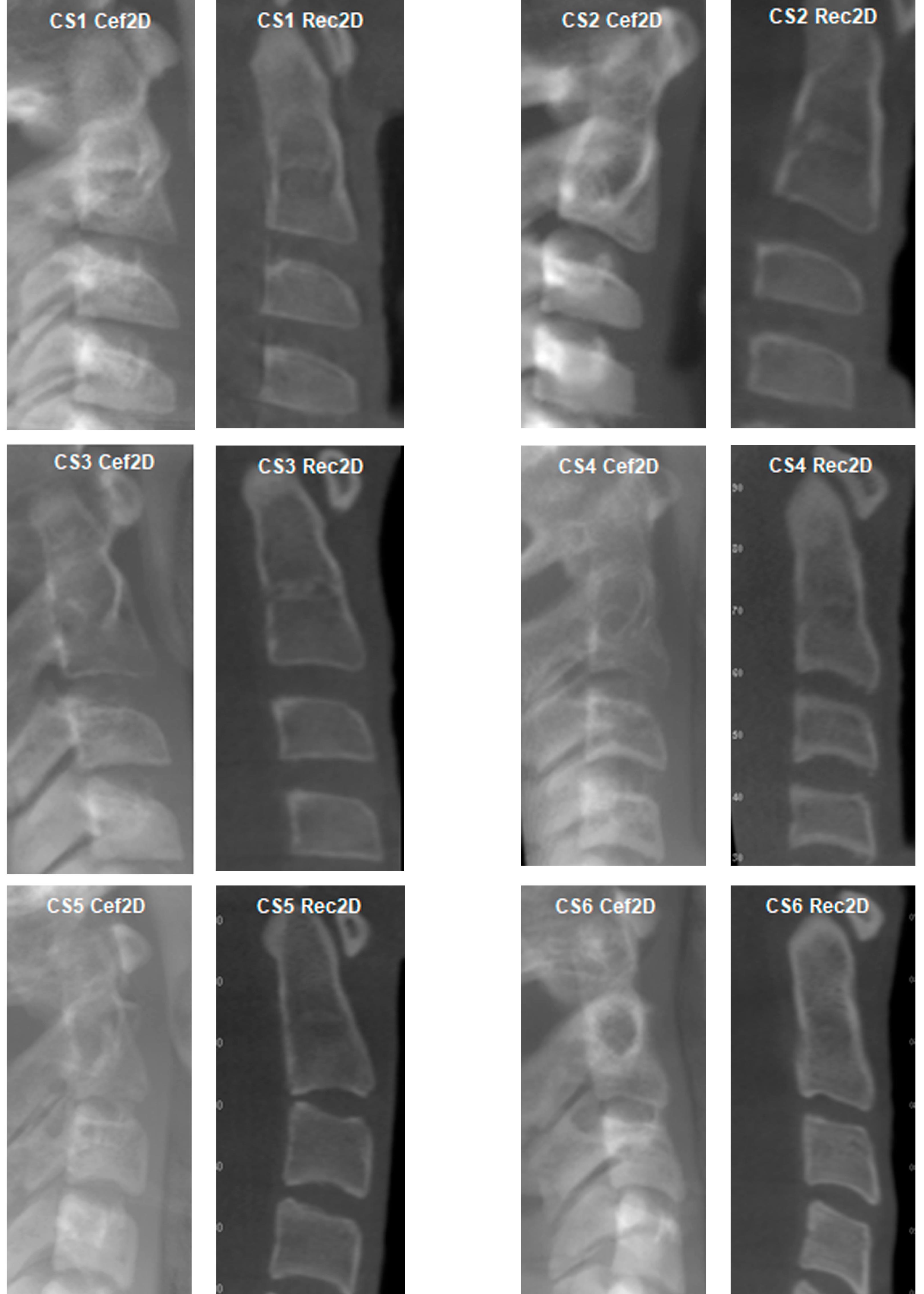
Figure 1.
Two-dimensional Cef2D and Rec2D images representing cervical vertebrae maturation stages (CS1–CS6) according to the CVM method proposed by Baccetti et al. (2005)
.
Two-dimensional Cef2D and Rec2D images representing cervical vertebrae maturation stages (CS1–CS6) according to the CVM method proposed by Baccetti et al. (2005)
For each image, all the participants completed evaluation forms, answering the following questions regarding the morphology of the C2, C3, and C4 vertebrae: 1) whether the inferior borders of C2, C3, and C4 were straight or concave; 2) whether the shapes of the C3 and C4 cervical vertebrae were trapezoidal, horizontal rectangular, quadrangular, or vertical rectangular; 3) whether the stage of maturation of the cervical vertebrae would be classified as CS1, CS2, CS3, CS4, CS5, or CS6.
Before the first evaluation period (T1), a theoretical presentation was provided in the form of a video explaining the CVM method and offering instructions for completing the evaluation forms. To support participants during the analyses, a flyer was distributed containing graphic representations of the cervical vertebrae maturation stages.6
All the images were presented for evaluation in random order and projected individually. Each participant answered the aforementioned questions regarding the morphology and maturation stage of the cervical vertebrae for both Cef2D and Rec2D images.
Four weeks after T1, the second evaluation period (T2) was conducted. Under similar conditions, all the participants watched the instructional video on the CVM method again and repeated the process of analyzing the images presented during T1. The images were randomized again before T2, resulting in a total of 3,600 responses for the study (T1 + T2).
In investigating the parameters used as guidelines in the CVM method, this study aimed to answer the following questions: What is the degree of reproducibility in visualizing concavity on the inferior border of the cervical vertebrae? What is the degree of reproducibility in evaluating the shape of the body of the cervical vertebrae? Regarding the classification of vertebral maturation stages proposed by Baccetti et al.,6 what is the degree of reproducibility of this method? Since orthopedic treatments are performed before and during the pubertal growth spurt,6,16,17 what is the degree of reproducibility of the CVM method when considering the grouping of post-peak mandibular growth stages?
Statistical Analysis
The variables analyzed were represented by absolute and relative frequencies. Cohen’s kappa coefficient was used to assess intra- and inter-examiner reproducibility, based on the following agreement parameters:
0: poor
0.01–0.20: weak
0.21–0.40: reasonable
0.41–0.60: moderate
0.61–0.80: substantial
0.81–1.00: almost perfect29
Statistical analysis was performed using SPSS 18.0 (SPSS Inc., Chicago, IL, USA), with a significance level set at 5% (P ≤ 0.05).
Results
Analysis of the data revealed divergences in the responses regarding the inferior borders of the C2, C3, and C4 vertebrae, the shape of the bodies of the C3 and C4 vertebrae, and their corresponding maturation stages.
The response combinations were categorized as either coherent or incoherent (Table 1). A coherent analysis was defined as a combination of responses for the inferior border and vertebral shape that matched the stages predicted by the CVM classification.6 This group comprised 2,718 responses across both time intervals (T1 and T2), representing 75.5% of the sample. An incoherent analysis arose from two situations: (I) when the combination of responses regarding the inferior border and vertebral shape did not correspond to any valid stage according to the CVM method (invalid stage); or (II) when the combination led to a valid stage, but the examiner incorrectly classified it (classification error). In total, 882 incoherent responses were identified across T1 and T2, representing 24.5% of the sample.
Table 1.
Coherences and incoherences identified in the evaluation of two-dimensional Cef2D and Rec2D images during T1 and T2 evaluation periods
|
|
|
|
Cef2D
|
Rec2D
|
|
|
|
|
n
|
%
|
n
|
%
|
| T1 |
Coherent |
|
704 |
78.2 |
623 |
69.2 |
|
|
Incoherent |
Invalid stage |
115 |
12.8 |
170 |
18.9 |
|
|
|
Classification error |
81 |
9 |
107 |
11.9 |
| T2 |
Coherent |
|
710 |
78.9 |
681 |
75.7 |
|
|
Incoherent |
Invalid stage |
116 |
12.9 |
149 |
16.6 |
|
|
|
Classification error |
74 |
8.2 |
70 |
7.8 |
n Cef2D = 900, n Rec2D = 900, for T1; n Cef2D = 900, n Rec2D = 900, for T2
For analyses concerning the inferior borders of C2, C3, and C4, and the shapes of C3 and C4, all the responses were considered. Regarding vertebral maturation stages based on the CVM method,6 three assessment models were established: (I) General CVM Stage—all responses were considered; (II) Coherent CVM Stage—only responses without inconsistencies; (III) Grouped Coherent CVM Stage—consistent responses in which post-mandibular growth peak stages were grouped.
Tables 2 and 3 present inter- and intra-examiner agreement levels, as expressed by Cohen’s kappa coefficient, respectively.29
Table 2.
Inter-examiner agreement for the variables used in the analysis of cervical vertebral maturation in two-dimensional Cef2D and Rec2D images during T1 evaluation period of the study
|
|
|
Inferior border**
|
Shape**
|
General CVM stage**
|
Coherent CVM stage***
|
Grouped coherent CVM stage***
|
| Cef2D |
Agreement n (%) |
798 (88.7) |
527 (58.6) |
632 (70.2) |
461 (76.3) |
555 (91.9) |
| Kappa (p)* |
0.83 |
0.454 |
0.643 |
0.714 |
0.882 |
| Agreement level |
Almost perfect |
Moderate |
Substantial |
Substantial |
Almost perfect |
| Rec2D |
Agreement n (%) |
810 (90.0) |
490 (54.4) |
688 (76.4) |
460 (86.6) |
509 (95.9) |
| Kappa (p)* |
0.85 |
0.441 |
0.718 |
0.836 |
0.927 |
| Agreement level |
Almost perfect |
Moderate |
Substantial |
Almost perfect |
Almost perfect |
*Kappa P < 0.001; **n Cef2D = 900, n Rec2D = 900; ***n Cef2D = 604, n Rec2D = 531
Table 3.
Intra-examiner agreement for the variables used in the analysis of cervical vertebral maturation in two-dimensional Cef2D and Rec2D images
|
|
|
Inferior border**
|
Shape**
|
General CVM stage**
|
Coherent CVM stage***
|
Grouped coherent CVM stage***
|
| Cef2D |
Agreement n (%) |
775 (86.1) |
525 (58,3) |
612 (68.0) |
464 (76.8) |
533 (88.2) |
| Kappa (p)* |
0.791 |
0.473 |
0.616 |
0.72 |
0.829 |
| Agreement level |
Substantial |
Moderate |
Substantial |
Substantial |
Almost perfect |
| Rec2D |
Agreement n (%) |
785 (87.2) |
533 (59.2) |
621 (69.1) |
443 (83.4) |
496 (93.4) |
| Kappa (p)* |
0.805 |
0.499 |
0.626 |
0.796 |
0.881 |
| Agreement level |
Substantial |
Moderate |
Substantial |
Substantial |
Almost perfect |
*Kappa P < 0.001; **n Cef2D = 900, n Rec2D = 900; ***n Cef2D = 604, n Rec2D = 531
Discussion
The use of three-dimensional (3D) technology has revolutionized diagnostic imaging methods in orthodontics;30 however, the assessment of skeletal development stages through the analysis of CVM in CBCT scans has not been adequately explored. Recent studies have shown that CBCT scans can be used as a valid and reliable diagnostic tool for this purpose, comparable to cephalometric radiographs.31,32 Field of view (FOV) options meet the requirements for CBCT examinations in pediatric orthodontic patients with various specific indications, including 3D cephalometric analyses.33 Customized optimization strategies can provide more accurate and effective treatment planning, achieving reduced radiation doses through improved accessibility and minimizing the need for additional 2D images.33‒35 In this context, the ALADAIP principle (“as low as diagnostically acceptable, being indication-oriented and patient-specific”) plays a crucial role in minimizing radiation exposure, as comprehensive imaging of the entire head is essential for orthodontic planning.36
The literature has reported controversies regarding the analysis of the morphological characteristics of cervical vertebrae, with studies revealing high reproducibility of the CVM method4,7,10,13,14 and levels of agreement ranging from almost perfect6,8,9,11,12,19 to substantial,15 while other studies reported low reproducibility.16-18,20,21 In the majority of previous studies, the degree of reproducibility of the CVM method evaluated exclusively the stage of maturation.9,11,15,16,19-21,26,37,38 However, it is important to consider the inclusion of morphological characteristics of cervical vertebrae and the classification of their stages, as performed by Nestman et al.17 and Sohrabi et al.18 Considering the inter-examiner assessment at T1 in the present study, the degree of reproducibility related to the inferior border of the vertebrae presented an almost perfect level of agreement (Table 2, Additional File 2), disagreeing with the moderate to substantial levels of agreement previously mentioned in the literature.17,18 However, regarding the shape, the level of agreement was moderate, which corroborates the findings of Sohrabi et al.,18 but differs from the reasonable level reported by Nestman et al17 (Table 2, Additional File 2). Regarding the intra-examiner assessment, the levels of agreement for the inferior border and the shape of the cervical vertebrae were substantial and moderate, respectively (Table 3, Additional File 3). In contrast, Sohrabi et al.18 found substantial to almost perfect levels of agreement for the inferior border and substantial levels of agreement for the shape of the cervical vertebrae.
To date, this is the first study in which participants themselves directly determined the CVM stage based on their evaluations of the inferior borders and shapes of the cervical vertebrae. When the inferior border and vertebral shape did not clearly correspond to a stage defined in the CVM method, the participants tended to rely more heavily on the inferior border when making their final determination of the maturation stage. This is a noteworthy distinction, as in the studies by Nestman et al.17 and Sohrabi et al.,18 the CVM stage was not directly defined by participants but was determined by the authors based on participants’ assessments of the inferior border and vertebral shape.
Out of the 3,600 responses in this study, 24.5% were classified as incoherent (Table 1, Additional File 1). A common error involved misclassifying responses that should correspond to stage CS5 as CS4. Similar rates of incoherent responses in CVM classification have been reported in the literature,17,21,39 ranging from 14% to 30%.17,39 In particular, frequent incoherencies have been noted in stage CS4,39 as well as the observation of horizontal rectangular shapes for C3 and C4 in some adult patients.21
Concerning the degree of reproducibility relative to the Coherent CVM Stage, the level of inter-examiner agreement was almost perfect for Rec2D and substantial for Cef2D (Table 2, Additional File 2). This was the only difference observed between the two image types concerning the reproducibility parameters evaluated. These findings demonstrate a higher degree of reproducibility than those reported in previous studies, which indicated moderate inter-examiner agreement.17,18
When comparing responses related to the inferior border between Cef2D and Rec2D for the same individuals, virtually no difference was observed in the present study. However, there was greater variability in shape-related combinations. Hassel and Farman reported that this could be associated with the presence of overlaps or noise in radiographic images, which reduces image clarity.4 The literature has consistently emphasized the importance of analyzing the inferior borders of the cervical vertebrae in relation to their maturation stages. San Román et al.40 evaluated the anatomical parameters of the inferior border, vertebral height, and shape, and verified that, when each of these parameters was individually compared with skeletal maturation based on hand and wrist radiographs, the concavity of the inferior border demonstrated the strongest correlation.
Clinically, in cases of sagittal and transverse maxillary deficiency, the most appropriate time for orthopedic intervention is during the pre-pubertal phase, specifically at stages CS1 and CS2. For cases involving mandibular ramus deficiency and mandibular retrognathism, the ideal time for achieving more effective orthopedic treatment corresponds to the peak growth period, represented by stage CS3.6,12,41,42 In the literature, stage CS4 is described as a pubertal stage.37,42 However, it is important to note that, according to Baccetti et al.,6 the mandibular growth peak occurs at least one to two years prior to this stage. Based on the results presented, although the analysis of the inferior borders of the cervical vertebrae demonstrated an almost perfect level of agreement, the evaluation of vertebral shape remains a controversial factor that may compromise the reproducibility of the CVM method. Therefore, it is suggested that only the presence or absence of concavity on the inferior borders of the C2, C3, and C4 vertebrae should be considered, while grouping the post-peak mandibular growth stages—CS4, CS5, and CS6—since changes occurring in these stages are primarily related to vertebral shape (Figure 2).
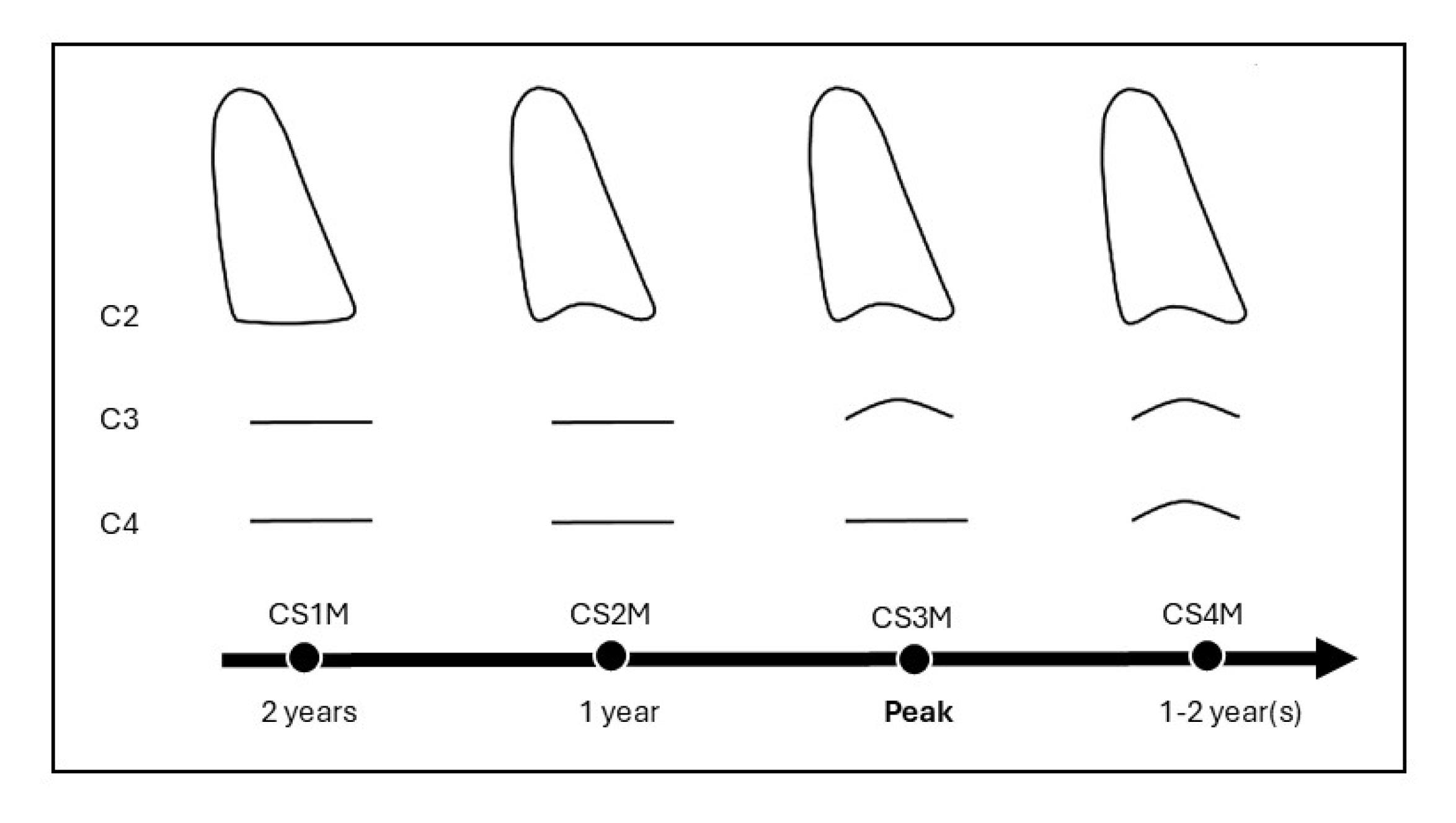
Figure 2.
Diagram of the CVM method recommended by Baccetti et al. (2005), illustrating the inferior borders of vertebrae C3 and C4. Modified cervical stage 1 (CS1M): The inferior borders of C2, C3, and C4 are flat. The mandibular growth peak will occur, on average, two years after this stage. Modified cervical stage 2 (CS2M): The inferior border of C2 is concave, while those of C3 and C4 remain flat. The mandibular growth peak will occur, on average, one year after this stage. Modified cervical stage 3 (CS3M): The inferior borders of C2 and C3 are concave, and that of C4 remains flat. The mandibular growth peak will occur during the year in which this stage is identified. Modified cervical stage 4 (CS4M): The inferior borders of C2, C3, and C4 are all concave. The mandibular growth peak occurred at least one to two years before this stage
.
Diagram of the CVM method recommended by Baccetti et al. (2005), illustrating the inferior borders of vertebrae C3 and C4. Modified cervical stage 1 (CS1M): The inferior borders of C2, C3, and C4 are flat. The mandibular growth peak will occur, on average, two years after this stage. Modified cervical stage 2 (CS2M): The inferior border of C2 is concave, while those of C3 and C4 remain flat. The mandibular growth peak will occur, on average, one year after this stage. Modified cervical stage 3 (CS3M): The inferior borders of C2 and C3 are concave, and that of C4 remains flat. The mandibular growth peak will occur during the year in which this stage is identified. Modified cervical stage 4 (CS4M): The inferior borders of C2, C3, and C4 are all concave. The mandibular growth peak occurred at least one to two years before this stage
Conclusion
The optimization of the cervical vertebral maturation (CVM) method proposed by Baccetti et al. (2005) for application in CBCT demonstrated a higher level of reproducibility, particularly when considering the inferior borders of the C2, C3, and C4 vertebrae, as well as the grouping of post-peak stages of mandibular growth.
Competing Interests
The authors declare no competing interests related to this research.
Ethical Approval
This study was approved by the Human Research Ethics Committee of the Clementino Fraga Filho Hospital, Universidade Federal do Rio de Janeiro, Brazil (CAAE 71244217.9.0000.5257, opinion number 2.203.180/2017). All the participants who agreed to take part in the study gave their informed consent before starting it.
Acknowledgements
The authors gratefully acknowledge the Postgraduate Program in Dentistry, the Department of Oral Diagnosis and Pathology, and the Department of Pediatric Dentistry and Orthodontics at the Universidade Federal do Rio de Janeiro, as well as all participants, for their valuable support and contributions.
References
- Fishman LS. Radiographic evaluation of skeletal maturation A clinically oriented method based on hand-wrist films. Angle Orthod 1982; 52(2):88-112. doi: 10.1043/0003-3219(1982)052<0088:Reosm>2.0.Co;2 [Crossref] [ Google Scholar]
- Soegiharto BM, Cunningham SJ, Moles DR. Skeletal maturation in Indonesian and white children assessed with hand-wrist and cervical vertebrae methods. Am J Orthod Dentofacial Orthop 2008; 134(2):217-26. doi: 10.1016/j.ajodo.2006.07.037 [Crossref] [ Google Scholar]
- Greulich WW, Pyle SI. Radiographic Atlas of Skeletal Development of the Hand and Wrist. 2nd ed. Stanford: Stanford University Press; 1959.
- Hassel B, Farman AG. Skeletal maturation evaluation using cervical vertebrae. Am J Orthod Dentofacial Orthop 1995; 107(1):58-66. doi: 10.1016/s0889-5406(95)70157-5 [Crossref] [ Google Scholar]
- Baccetti T, Franchi L, McNamara JA Jr. An improved version of the cervical vertebral maturation (CVM) method for the assessment of mandibular growth. Angle Orthod 2002; 72(4):316-23. doi: 10.1043/0003-3219(2002)072<0316:Aivotc>2.0.Co;2 [Crossref] [ Google Scholar]
- Baccetti T, Franchi L, McNamara JA Jr. The cervical vertebral maturation (CVM) method for the assessment of optimal treatment timing in dentofacial orthopedics. Semin Orthod 2005; 11(3):119-29. doi: 10.1053/j.sodo.2005.04.005 [Crossref] [ Google Scholar]
- Franchi L, Baccetti T, McNamara JA Jr. Mandibular growth as related to cervical vertebral maturation and body height. Am J Orthod Dentofacial Orthop 2000; 118(3):335-40. doi: 10.1067/mod.2000.107009 [Crossref] [ Google Scholar]
- Bonfim MA, Costa AL, Fuziy A, Ximenez ME, Cotrim-Ferreira FA, Ferreira-Santos RI. Cervical vertebrae maturation index estimates on cone beam CT: 3D reconstructions vs sagittal sections. Dentomaxillofac Radiol 2016; 45(1):20150162. doi: 10.1259/dmfr.20150162 [Crossref] [ Google Scholar]
- Pasciuti E, Franchi L, Baccetti T, Milani S, Farronato G. Comparison of three methods to assess individual skeletal maturity. J Orofac Orthop 2013; 74(5):397-408. doi: 10.1007/s00056-013-0164-x [Crossref] [ Google Scholar]
- Soegiharto BM, Moles DR, Cunningham SJ. Discriminatory ability of the skeletal maturation index and the cervical vertebrae maturation index in detecting peak pubertal growth in Indonesian and white subjects with receiver operating characteristics analysis. Am J Orthod Dentofacial Orthop 2008; 134(2):227-37. doi: 10.1016/j.ajodo.2006.09.062 [Crossref] [ Google Scholar]
- Uysal T, Sari Z, Ramoglu SI, Basciftci FA. Relationships between dental and skeletal maturity in Turkish subjects. Angle Orthod 2004; 74(5):657-64. doi: 10.1043/0003-3219(2004)074<0657:Rbdasm>2.0.Co;2 [Crossref] [ Google Scholar]
- Goyal S, Goyal S. Assessment of skeletal maturation and pubertal growth spurt using cervical vertebrae maturation indicators. Rwanda Med J 2013; 70(3):28-33. [ Google Scholar]
- Beit P, Peltomäki T, Schätzle M, Signorelli L, Patcas R. Evaluating the agreement of skeletal age assessment based on hand-wrist and cervical vertebrae radiography. Am J Orthod Dentofacial Orthop 2013; 144(6):838-47. doi: 10.1016/j.ajodo.2013.07.015 [Crossref] [ Google Scholar]
- Ozer T, Kama JD, Yiğit Özer S. A practical method for determining pubertal growth spurt. Am J Orthod Dentofacial Orthop 2006;130(2):131.e1-6. doi: 10.1016/j.ajodo.2006.01.019.
- Rainey BJ, Burnside G, Harrison JE. Reliability of cervical vertebral maturation staging. Am J Orthod Dentofacial Orthop 2016; 150(1):98-104. doi: 10.1016/j.ajodo.2015.12.013 [Crossref] [ Google Scholar]
-
Gabriel DB, Southard KA, Qian F, Marshall SD, Franciscus RG, Southard TE. Cervical vertebrae maturation method: poor reproducibility. Am J Orthod Dentofacial Orthop 2009;136(4):478.e1-7. doi: 10.1016/j.ajodo.2007.08.028.
- Nestman TS, Marshall SD, Qian F, Holton N, Franciscus RG, Southard TE. Cervical vertebrae maturation method morphologic criteria: poor reproducibility. Am J Orthod Dentofacial Orthop 2011; 140(2):182-8. doi: 10.1016/j.ajodo.2011.04.013 [Crossref] [ Google Scholar]
- Sohrabi A, Babay Ahari S, Moslemzadeh H, Rafighi A, Aghazadeh Z. The reliability of clinical decisions based on the cervical vertebrae maturation staging method. Eur J Orthod 2016; 38(1):8-12. doi: 10.1093/ejo/cjv030 [Crossref] [ Google Scholar]
- Maló L, Lima S, Teixeira V, Canova F, Alves S. Skeletal maturation in a Portuguese population – comparison between the hand‐wrist and cervical vertebral maturation. Rev Port Estomatol Med Dent Cir Maxilofac 2014; 55(2):102-9. doi: 10.1016/j.rpemd.2014.05.001 [Crossref] [ Google Scholar]
- Predko-Engel A, Kaminek M, Langova K, Kowalski P, Fudalej PS. Reliability of the cervical vertebrae maturation (CVM) method. Bratisl Lek Listy 2015; 116(4):222-6. doi: 10.4149/bll_2015_043 [Crossref] [ Google Scholar]
- Zhao XG, Lin J, Jiang JH, Wang Q, Ng SH. Validity and reliability of a method for assessment of cervical vertebral maturation. Angle Orthod 2012; 82(2):229-34. doi: 10.2319/051511-333.1 [Crossref] [ Google Scholar]
- Joshi V, Yamaguchi T, Matsuda Y, Kaneko N, Maki K, Okano T. Skeletal maturity assessment with the use of cone-beam computerized tomography. Oral Surg Oral Med Oral Pathol Oral Radiol 2012; 113(6):841-9. doi: 10.1016/j.oooo.2011.11.018 [Crossref] [ Google Scholar]
- Byun BR, Kim YI, Yamaguchi T, Maki K, Son WS. Quantitative assessment of cervical vertebral maturation using cone beam computed tomography in Korean girls. Comput Math Methods Med 2015; 2015:405912. doi: 10.1155/2015/405912 [Crossref] [ Google Scholar]
- Angelieri F, Franchi L, Cevidanes LH, McNamara JA Jr. Diagnostic performance of skeletal maturity for the assessment of midpalatal suture maturation. Am J Orthod Dentofacial Orthop 2015; 148(6):1010-6. doi: 10.1016/j.ajodo.2015.06.016 [Crossref] [ Google Scholar]
- Bassed RB, Briggs C, Drummer OH. Analysis of time of closure of the spheno-occipital synchondrosis using computed tomography. Forensic Sci Int 2010; 200(1-3):161-4. doi: 10.1016/j.forsciint.2010.04.009 [Crossref] [ Google Scholar]
- Shim JJ, Heo G, Lagravère MO. Assessment of skeletal maturation based on cervical vertebrae in CBCT. Int Orthod 2012; 10(4):351-62. doi: 10.1016/j.ortho.2012.09.005 [Crossref] [ Google Scholar]
- Abramson JH. WINPEPI updated: computer programs for epidemiologists, and their teaching potential. Epidemiol Perspect Innov 2011; 8(1):1. doi: 10.1186/1742-5573-8-1 [Crossref] [ Google Scholar]
- de Oliveira Ruellas AC, Tonello C, Gomes LR, Yatabe MS, Macron L, Lopinto J. Common 3-dimensional coordinate system for assessment of directional changes. Am J Orthod Dentofacial Orthop 2016; 149(5):645-56. doi: 10.1016/j.ajodo.2015.10.021 [Crossref] [ Google Scholar]
- Landis JR, Koch GG. The measurement of observer agreement for categorical data. Biometrics 1977; 33(1):159-74. doi: 10.2307/2529310 [Crossref] [ Google Scholar]
- Polizzi A, Serra S, Leonardi R. Use of CBCT in orthodontics: a scoping review. J Clin Med 2024; 13(22):6941. doi: 10.3390/jcm13226941 [Crossref] [ Google Scholar]
- Tekın A, Cesur Aydın K. Comparative determination of skeletal maturity by hand-wrist radiograph, cephalometric radiograph and cone beam computed tomography. Oral Radiol 2020; 36(4):327-36. doi: 10.1007/s11282-019-00408-y [Crossref] [ Google Scholar]
- Echevarría-Sánchez G, Arriola-Guillén LE, Malpartida-Carrillo V, Tinedo-López PL, Palti-Menendez R, Guerrero ME. Reliability of cephalograms derived of cone beam computed tomography versus lateral cephalograms to estimate cervical vertebrae maturity in a Peruvian population: a retrospective study. Int Orthod 2020; 18(2):258-65. doi: 10.1016/j.ortho.2020.01.001 [Crossref] [ Google Scholar]
- Kissel P, Mah JK, Bumann A. Modern 3D cephalometry in pediatric orthodontics-downsizing the FOV and development of a new 3D cephalometric analysis within a minimized large FOV for dose reduction. Clin Oral Investig 2021; 25(7):4651-70. doi: 10.1007/s00784-021-03779-x [Crossref] [ Google Scholar]
- Horner K. Cone Beam CT for Dental and Maxillofacial Radiology (Evidence Based Guidelines). Luxembourg: European Commission, Directorate-General for Energy; 2012. Available from: http://ec.europa.eu/energy/nuclear/radiation_protection/doc/publication/172.pdf.
- Oliveira PL, Starling CR, Maurício CL, Guedes FR, Visconti MA, Ruellas AC. Ionizing radiation and its considerations in imaging diagnosis: comparison of absorbed dose between cone beam computed tomography and multi-detector computed tomography in the head and neck. Radioprotection 2021; 56(2):111-5. doi: 10.1051/radiopro/2021006 [Crossref] [ Google Scholar]
- Oenning AC, Jacobs R, Salmon B. ALADAIP, beyond ALARA and towards personalized optimization for paediatric cone-beam CT. Int J Paediatr Dent 2021; 31(5):676-8. doi: 10.1111/ipd.12797 [Crossref] [ Google Scholar]
- Perinetti G, Caprioglio A, Contardo L. Visual assessment of the cervical vertebral maturation stages: a study of diagnostic accuracy and repeatability. Angle Orthod 2014; 84(6):951-6. doi: 10.2319/120913-906.1 [Crossref] [ Google Scholar]
- Santos EC, Bertoz FA, de Moraes Arantes F, Reis PM. Evaluation of the reproducibility of the method of determination of the skeletal maturation by cervical vertebrae. Rev Dent Press Ortodon Ortop Facial 2005; 10(2):62-8. doi: 10.1590/s1415-54192005000200009 [Crossref] [ Google Scholar]
- Perinetti G, Bianchet A, Franchi L, Contardo L. Cervical vertebral maturation: an objective and transparent code staging system applied to a 6-year longitudinal investigation. Am J Orthod Dentofacial Orthop 2017; 151(5):898-906. doi: 10.1016/j.ajodo.2016.09.026 [Crossref] [ Google Scholar]
- San Román P, Palma JC, Oteo MD, Nevado E. Skeletal maturation determined by cervical vertebrae development. Eur J Orthod 2002; 24(3):303-11. doi: 10.1093/ejo/24.3.303 [Crossref] [ Google Scholar]
- Mohammad Z, Namineni S, Cheruku SR, Penmetcha S, Munnangi SR, Mallineni SK. Evaluation of relationship of hand wrist radiograph, cervical vertebral maturation with mandibular growth as skeletal maturity indicators in children. European J Gen Dent 2016; 5(1):24-9. doi: 10.4103/2278-9626.172737 [Crossref] [ Google Scholar]
- Perinetti G, Franchi L, Contardo L. Determination of timing of functional and interceptive orthodontic treatment: a critical approach to growth indicators. J World Fed Orthod 2017; 6(3):93-7. doi: 10.1016/j.ejwf.2017.08.006 [Crossref] [ Google Scholar]