J Dent Res Dent Clin Dent Prospects. 20(1):56-63.
doi: 10.34172/joddd.42747
Original Article
Effect of Resin, Calcium Silicate, and Silicone-Based Root Canal Sealers on Postoperative Pain: A Randomized Controlled Clinical Trial
Gizem Kadı Conceptualization, Data curation, Formal analysis, Investigation, Methodology, Supervision, Validation, Writing – original draft, Writing – review & editing, 1, * 
Esin Özlek Formal analysis, Investigation, Methodology, Project administration, Supervision, Validation, Writing – original draft, Writing – review & editing, 2
Author information:
1Department of Endodontology, Faculty of Dentistry, Altınbas University, İstanbul, Turkey
2Department of Endodontology, Faculty of Dentistry, Van Yuzuncu Yil University, Van, Turkey
Abstract
Introduction:
This study aimed to compare the effects of resin- (AH Plus), calcium silicate- (iRoot SP), and silicone-based (GuttaFlow Bioseal) root canal sealers on postoperative pain.
Methods:
This study included 84 patients with mandibular molars diagnosed with irreversible pulpitis. Root canal shaping procedures were completed using X-Smart Plus (Dentsply Maillefer, Ballaigues, Switzerland) endodontic motor and EndoArt (İnci Dental, İstanbul, Turkey) nickel-titanium rotary system up to #25 (30.06) shaping file for mesial root anals and #30 (0.6) shaping file for root distal canals. The patients were randomly divided into 3 groups based on the root canal sealer used (n=28): group 1: AH Plus, group 2: iRoot SP, and group 3: GuttaFlow Bioseal. Pain scores were recorded using a visual analog scale (VAS) before and after treatment at the 6th, 24th, 48th, and 72nd hours, and at the first week. Additionally, the frequency and timing of analgesic intake were recorded. The chi-squared test was used for categorical variables. The Kruskal-Wallis test was used to compare differences in non-normally distributed continuous data between groups. The Friedman test was used to compare pain scores over time within each group, and the Cochran’s Q test was used to analyze analgesic use.
Results:
No statistically significant differences in median VAS scores were found at various time intervals between the groups (P>0.05). Analgesic use distribution at the 6th (P=0.658) and the 24th (P=0.355) hours also did not show significant differences between the groups.
Conclusion:
Resin-, bioceramic-, or silicone-based sealers showed similar effects on postoperative pain.
Keywords: Clinical trial, Postoperative pain, Root canal sealer
Copyright and License Information
© 2026 The Author(s).
This is an open access article distributed under the terms of the Creative Commons Attribution License (
http://creativecommons.org/licenses/by/4.0/), which permits unrestricted use, distribution, and reproduction in any medium, provided the original work is properly cited.
Funding Statement
This research received no specific grant from any funding agency in the public, commercial, or not-for-profit sectors.
Introduction
Postoperative pain, which starts within hours or days following endodontic treatment, is an undesirable condition for both the patient and clinician. The etiology of postoperative pain is multifactorial and is affected by factors related to the patient (age, gender, general health status, and psychological factors), tooth (tooth type, pulp and periradicular tissue status, presence of fistula, and presence of preoperative pain) and treatment procedures (length of operation, instrumentation, irrigation, obturation, number of sessions, and occlusal reduction)1,2 with reported incidence rates ranging from 3% to 58%.3 Among the predisposing factors are incomplete cleaning and shaping or obturation procedures.4 Root canal sealers can penetrate periapical tissues through apical foramina and lateral canals, or due to apical extrusion. Consequently, they may cause cytotoxic effects on periapical tissues, leading to inflammation, necrosis, and postoperative pain.5
The epoxy-resin-based root canal sealer AH Plus (Dentsply, DeTrey, Konstanz, Germany) is widely used due to its low solubility, strong apical sealing, and satisfactory dentin adhesion.6 Recently, bioceramic-based root canal sealers have gained popularity as bioactive materials with less cytotoxicity compared to resin-based sealers. iRoot SP (BioCeramix, Vancouver, Canada), a bioceramic-based root canal sealer containing calcium silicate phosphate, is noted for its biocompatibility, superior physical properties, and the ability to maintain alkalinity for 14 days due to its calcium ion release properties.7–9
GuttaFlow Bioseal (Coltene, Whaledent, Langenau, Switzerland) is a silicone-based root canal sealer with added calcium silicate particles. It is also a bioactive glass, with a finely ground gutta-percha powder and silicone-based matrix that disperses homogeneously upon mixing.10,11 It has a high water-absorption capacity, low solubility, and porosity. Additionally, it offers the benefits of alkalinization through calcium ion release and the formation of hydroxyapatite.12,13 Studies have reported that GuttaFlow Bioseal expands more than AH Plus after hardening, resulting in less leakage in root canal fillings and lower cytotoxicity.10,14,15
Although several studies in the literature have focused on postoperative pain and its association with etiological factors and root canal sealers, to the best of our knowledge, no study has examined the effects of GuttaFlow Bioseal root canal sealer on postoperative pain. This study investigated and compared the effects of single-visit root canal treatments with AH Plus, iRoot SP, and GuttaFlow Bioseal sealers on postoperative pain 6, 24, 48, and 72 hours and one week after treatment. The null hypotheses were that there is no difference in the incidence and intensity of post-obturation pain among the AH plus, iRoot SP, and GuttaFlow Bioseal groups following single-visit root canal treatment.
Methods
Study Design, Setting, and Sampling
This randomized clinical trial was performed according to the Preferred Reporting Items for Randomized Trials in Endodontics (PRIRATE) 2020 guidelines. Ethical approval for the study was obtained from the Institutional Review Board and the Ethics Committee of Van Yuzuncu Yıl University (19/29.04.2021). The Consolidated Standards of Reporting Trials (CONSORT) guidelines were followed in this clinical trial, and the study protocol was registered on www.clinicaltrial.gov (Identifier: NCT05033093) and first posted on 26/08/2021. Voluntary patients were included in this study. All the patients signed an informed consent form after the objectives, procedures, benefits, and potential risks of the study were explained. The Power and Sample Size Calculation software version 3.1.2 was used to calculate the sample size.16 With 95% confidence, 80% test power, and f = 0.3476158 impact size, the total sample size was determined at n = 84. A total of 84 patients (28 per group) underwent single-visit root canal treatment performed by the same dentist.
Patient Selection and Pretreatment Assessment
Healthy individuals aged 18 to 60, without systemic diseases, presenting with mandibular molar teeth exhibiting asymptomatic pulpitis perforating the vital pulp and requiring root canal treatment for preprosthetic purposes were eligible for inclusion. Additionally, teeth demonstrating positive responses to thermal and electric pulp tests were considered.
Exclusion criteria included individuals who had used antibiotics, painkillers, or anti-inflammatory medications in the past seven days, pregnant or breastfeeding individuals, those with traumatic malocclusion or occlusal contact issues, teeth with visible periapical lesions on radiographs, material loss preventing the use of a rubber dam, teeth showing resorption, open apices, or calcification, teeth with gingival recession > 3 mm, deep pockets, mobility, or roots that were fractured or cracked.
Patients underwent clinical examination, and periapical radiographs were obtained with the bisecting-angle technique. The gender (male/female), age, and preoperative pain scores of the patients were recorded. Preoperative pain assessment was conducted with the dentist to assess patient comprehension and cooperation with the pain scale. Patients unable to adequately engage with the scale were excluded from the study. Only one tooth per patient was included, and pulp vitality was assessed using an electric pulp testing device (Analytic Technology Corp., Redmond, WA, USA).
Randomization and Blinding
Patient allocation to study groups was achieved using the envelope method, ensuring randomization regardless of age and gender, in a 1:1 ratio. The participating patients were blinded to their assigned groups as they received no information regarding group allocation. However, due to the study design, the clinician could not be blinded.
Clinical Interventions
Local anesthesia was administered via inferior alveolar nerve block and buccal infiltration anesthesia using 2 mL of articaine hydrochloride containing 1:100,000 adrenaline (Ultraver DS fort; Haver, Istanbul, Turkey). Following rubber dam isolation, access cavities were prepared using a sterile round diamond bur. Tooth vitality was confirmed by observing bleeding during cavity preparation; teeth without bleeding were treated but excluded from the study. Working length (WL) was determined with an apex locator (Propex Pixi, Dentsply Maillefer) and confirmed to be 0.5‒1 mm shorter than the radiographic apex.
The root canals were shaped using the crown-down technique with an X-Smart Plus (Dentsply Maillefer, Ballaigues, Switzerland) endodontic motor and EndoArt (Inci Dental, Istanbul, Turkey) nickel-titanium rotary system. In line with the manufacturer’s recommendation, all root canals were shaped with an entry file at 300 rpm and 3.5 Ncm torque, and with shaping files (#15 (0.4) and #20 (0.4)) at 300 rpm and 1.5 Nm torque, respectively. Then, the shaping procedure of mesial root canals was completed with finishing files #25 (0.6) at 300 rpm and 2.5 Ncm torque. In addition to mesial root canals, the distal root canals were shaped with finishing file #30 (0.6) at the same speed and torque settings. During shaping, when resistance was encountered, the files were removed from the root canal to prevent excessive torsion. The apical opening was checked with a #10K file, and root canal preparation was continued. These interventions were repeated until the targeted working length was achieved.
During the shaping process, at each file change, root canal irrigation was performed with 2 mL of 5.25% NaOCl solution (Microvem AF, Istanbul, Turkey) for 1 minute. For the final irrigation of the root canals following the shaping process, 5.25% NaOCl, 17% EDTA (Imircryl, Konya, Turkey), 5.25% NaOCL, and saline solutions (5 mL of each) were used, respectively. For all irrigation applications, a 2-mL dental syringe and a #27G dental needle were used, both adjusted to be 2 mm shorter than the working length. After the final irrigation procedure, the root canals were dried with sterile paper points of matching diameter. Subsequently, the participants were categorized into three groups based on the type of root canal sealer used:
Group 1: AH Plus root canal sealer
Group 2: iRoot SP root canal sealer
Group 3: GuttaFlow Bioseal root canal sealer
After obtaining radiographic control images with a master cone matching the last used file at working length for the single-cone technique, the selected gutta-percha cone (EndoArt, Inci Dental, Istanbul, Turkey) was coated with root canal sealer and inserted into the root canal. The root canal was then filled with the root canal sealer at the working length using back-and-forth movements. Following the application of glass-ionomer filling material (Ionoseal, VOCO, Cuxhaven, IOS) to cover the root canal opening as a standard procedure, permanent restoration was completed with a single-stage, self-adhesive, light-activated composite resin (Filtek Z250, 3M ESPE, St. Paul, Minnesota, USA). The root canal fillings were evaluated using radiography. All the patients were prescribed 400 mg of ibuprofen, with instructions to use it only in cases of severe pain.
Postoperative Pain Evaluation
Postoperative pain was assessed using a visual analog scale (VAS).9 The patients were asked to record their perceived pain on the VAS at 6, 24, 48, and 72 hours, as well as one week after the procedure. Additionally, the patients were asked to record the number of analgesics used and the time of administration on the VAS scale. One week after the intervention, the patients were contacted by phone to obtain their recorded pain scores, which were then documented in the patient files. Patients who could not be reached by phone were excluded from the study.
Statistical Analysis
The statistical analysis of the data was performed using SPSS 23. Normal distribution of the data was assessed using the Kolmogorov-Smirnov test. Categorical variables were compared between groups using the chi-squared test. The Kruskal-Wallis test was used to compare the data, which were not normally distributed, across the groups. The Mann-Whitney U test was used to compare the pain scores by gender. The Friedman test was used to compare intergroup pain scores over time. The Cochran’s Q test was used to compare analgesic use. The Spearman’s Rho Correlation Coefficient was used to assess the correlation between age and pain scores. Regarding the analysis results, quantitative data were presented as mean ± standard deviation and median (minimum-maximum), and categorical data were presented as frequencies (percentages). The results were evaluated at a significance level of P < 0.050.
Results
Of the invited patients, 8 were excluded according to the established exclusion criteria, and 3 declined to participate. Consequently, they were excluded from the final analysis, leaving 84 participants (Figure 1). There were no statistically significant differences between the groups in gender (P = 0.953), age (P = 0.193), or preoperative pain scores (P = 0.766) (Table 1).
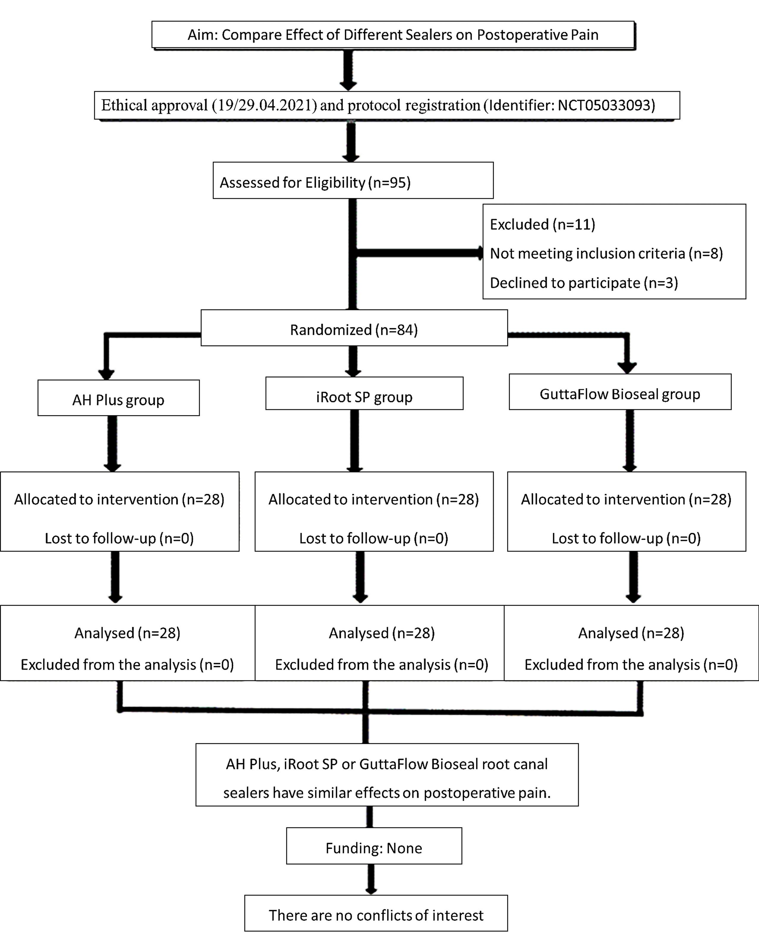
Figure 1.
PRIRATE 2020 flowchart showing the flow of participants in the study
.
PRIRATE 2020 flowchart showing the flow of participants in the study
Table 1.
Demographic and clinical features of the patients
|
|
AH Plus
|
iRoot SP
|
GuttaFlowBioseal
|
Total
|
Statistical Test
|
p
|
| Gender n (%) |
|
|
|
|
|
|
| Female |
15 (53.6) |
16 (57.1) |
16 (57.1) |
47 (56) |
χ2 = 0.097b |
0.953 |
| Male |
13 (46.4) |
12 (42.9) |
12 (42.9) |
37 (44) |
| Age |
27.57 ± 8.77 |
29.54 ± 7.33 |
31.68 ± 9.52 |
29.60 ± 8.65 |
χ2 = 3.295a |
0.193 |
| 24.50 (18.00‒51.00) |
28.50 (18.00‒46.00) |
31.50 (18.00‒57.00) |
29.00 (18.00‒57.00) |
| Preoperative pain |
2.04 ± 2.55 |
2.07 ± 2.48 |
2.04 ± 1.93 |
2.05 ± 2.31 |
χ2 = 0.532a |
0.766 |
| 1.00 (0.00‒8.00) |
0.50 (0.00‒7.00) |
2.00 (0.00‒7.00) |
1.00 (0.00‒8.00) |
aKruskal-Wallis test statistic, bchi-squared test statistic, mean ± standard deviation, median (min – max), frequency (percentage)
The effects of AH Plus, iRoot SP, and GuttaFlow Bioseal root canal sealers on postoperative pain were not statistically significant (P > 0.05). Regarding the intragroup comparison of different times, the most severe postoperative pain was observed at 6 hours, and there was a statistically significant difference compared to other times (P = 0.001) (Table 2).
Table 2.
Comparison of pain VAS scores between and within groups
|
|
AH Plus
|
iRoot SP
|
GuttaFlowBioseal
|
Total
|
Statistical Testb
|
p
|
| Preoperative pain score |
2.04 ± 2.55 |
2.07 ± 2.48 |
2.04 ± 1.93 |
2.05 ± 2.31 |
χ2 = 0.532 |
0.766 |
| 1.00 (0.00‒8.00)bc |
0.50 (0.00‒7.00)cd |
2.00 (0.00‒7.00)b |
1.00 (0.00‒8.00) |
| 6 hrs pain score |
2.86 ± 2.92 |
2.68 ± 2.39 |
2.04 ± 1.60 |
2.52 ± 2.36 |
χ2 = 0.736 |
0.692 |
| 2.50 (0.00‒10.00)b |
2.00 (0.00‒10.00)c |
2.00 (0.00‒6.00)b |
2.00 (0.00‒10.00) |
| 24 hrs pain score |
1.43 ± 2.15 |
1.43 ± 1.87 |
0.89 ± 1.40 |
1.25 ± 1.83 |
χ2 = 1.317 |
0.518 |
| 0.00 (0.00‒8.00)ab |
1.00 (0.00‒6.00)bc |
0.00 (0.00‒5.00)ab |
0.00 (0.00‒8.00) |
| 48 hrs pain score |
0.46 ± 1.29 |
0.61 ± 0.92 |
0.50 ± 1.17 |
0.52 ± 1.12 |
χ2 = 4.619 |
0.099 |
| 0.00 (0.00‒5.00)ac |
0.00 (0.00‒4.00)abd |
0.00 (0.00‒4.00)a |
0.00 (0.00‒5.00) |
| 72 hrs pain score |
0.21 ± 0.83 |
0.07 ± 0.38 |
0.11 ± 0.42 |
0.13 ± 0.58 |
χ2 = 0.423 |
0.809 |
| 0.00 (0.00‒4.00)a |
0.00 (0.00‒2.00)ab |
0.00 (0.00‒2.00)a |
0.00 (0.00‒4.00) |
| 1 week pain score |
0.00 ± 0.00 |
0.00 ± 0.00 |
0.00 ± 0.00 |
0.00 ± 0.00 |
--- |
--- |
| 0.00 (0.00‒0.00)a |
0.00 (0.00‒0.00)a |
0.00 (0.00‒0.00)a |
0.00 (0.00‒0.00) |
| Statistical Testa |
χ2 = 68.959 |
χ2 = 68.151 |
χ2 = 72.533 |
|
|
|
| pa |
< 0.001 |
< 0.001 |
< 0.001 |
|
|
|
a χ2: Friedman test statistic, b χ: Kruskal Wallis test statistic, a-d: No difference between times with the same letter within groups, mean ± standard deviation, median (min – max)
There was no statistically significant difference in analgesic use between the AH Plus, iRoot SP, and GuttaFlow Bioseal groups at different time intervals (6th, 24th, 48th, 72nd hours, and one week) (P > 0.05) (Table 3). Although there was no statistically significant difference in analgesic use across time points in the iRoot SP group, the highest incidence of analgesic use was observed at the 6th hour in the AH Plus and GuttaFlow Bioseal groups, with a statistically significant difference (P < 0.05).
Table 3.
Comparison of the use of analgesics between and within groups
|
|
AH Plus
|
iRoot SP
|
GuttaFlowBioseal
|
Total
|
Statistical Test
|
Pb
|
| 6 hrs analgesic intake |
|
|
|
|
|
|
| No |
23 (82.1)a |
25 (89.3) |
25 (89.3) |
73 (86.9) |
χ2 = 0.837 |
0.658 |
| Yes |
5 (17.9) |
3 (10.7) |
3 (10.7) |
11 (13.1) |
| 24 hrs analgesic intake |
|
|
|
|
|
|
| No |
26 (92.9)ab |
27 (96.4) |
28 (100) |
81 (96.4) |
χ2 = 2.074 |
0.355 |
| Yes |
2 (7.1) |
1 (3.6) |
0 (0) |
3 (3.6) |
| 48 hrs analgesic intake |
|
|
|
|
|
|
| No |
28 (100)b |
28 (100) |
28 (100) |
84 (100) |
--- |
--- |
| 72 hrs analgesic intake |
|
|
|
|
|
|
| No |
28 (100)b |
28 (100) |
28 (100) |
84 (100) |
--- |
--- |
| 1 week analgesic intake |
|
|
|
|
|
|
| No |
28 (100)b |
28 (100) |
28 (100) |
84 (100) |
--- |
--- |
| Test statistic |
Q = 16.0 |
Q = 8.5 |
Q = 12.0 |
|
|
|
| Pa |
0.003 |
0.075 |
0.017 |
|
|
|
χ: chi-squared test statistic, Q = Cochran’s Q test statistic, a-b: There is no difference between tenses with the same letter.
Bold numbers are P values for Cochran’s Q test
Discussion
Postoperative pain following root canal treatment is commonly attributed to acute inflammation in periapical tissues.17 The success of the root canal treatment depends not only on the efficacy of the clinical procedures or biological outcomes but also on the optimization of the patient comfort by minimizing postoperative pain.18 Consequently, this study aimed to assess the impact of three different root canal sealers (AH Plus, iRoot SP, and GuttaFlow Bioseal) used in the endodontic treatment on postoperative pain. According to the findings, the effects of AH Plus, iRoot SP, and GuttaFlow Bioseal root canal sealers on postoperative pain were not statistically significant, and the null hypothesis was accepted.
Epoxy-resin-based root canal sealers are associated with significant cytotoxic effects on periradicular tissues by inducing inflammatory mediators, including cyclooxygenase-2, nitric oxide synthase, receptor activator of nuclear factor kappa B ligand, and reactive oxygen species.19,20 Additionally, AH Plus has been linked to the release of monomers, such as bisphenol A and diglycidyl ether, which contribute to its cytotoxicity.21 Moreover, it has been reported to increase the expression of pro-inflammatory cytokines such as interleukin-6 and interleukin-8, and to significantly decrease mitochondrial activity.22 The literature contains only a limited number of studies on the effects of bioceramic-based root canal sealers on postoperative pain. To our knowledge, no other study has investigated the effect of silicon-based root canal sealers on postoperative pain, preventing direct comparison of our findings. Studies have shown that AH Plus exhibits higher cytotoxicity than both bioceramic-based8 and silicone-based14,23 root canal sealers, and causes more neurotoxic effects.24 Consistent with our findings, Ateş et al.25 reported that AH Plus caused more postoperative pain than iRoot SP; Aslan and Donmez26 found that AH Plus resulted in more postoperative pain than EndoSequence BC Sealer; Tan et al.27 reported that AH Plus led to more postoperative pain than Total Fill; Shim et al.28 indicated that AH Plus caused more postoperative pain than Endoseal MTA; and Ferreira et al.29 reported that AH Plus caused more postoperative pain than MTA Fillapex and Endofill.
The physical and chemical properties of root canal sealers, such as pH and consistency, are considered crucial factors influencing the severity of postoperative endodontic pain.30,31 The alkaline environment caused by root canal sealers plays a vital role in periapical recovery by inhibiting osteoclastic activity and promoting hard tissue formation through alkaline phosphatase activation.32 It was reported that in 7 days, the pH increased up to 8.1732, 8.7530, and 9.2112 with AH Plus, iRoot SP, and GuttaFlow Bioseal, respectively. It was reported that GuttaFlow Bioseal released 3.80 mg/L Ca2+ions at the end of the 14th day, which increased to 3.89 mg/L at the end of the 28th day.9 The Ca2+ion release increased to 2.96 mg/L within 14 days, but this level was not sustained at the end of the 28th day and dropped to 2.015 mg/L.30 In contrast, AH Plus root canal sealer exhibited significantly less Ca2+ion release ( < 1.00) compared to other sealers.31,33 Although there was no statistically significant difference in postoperative pain between the root canal sealers preferred in this study, AH Plus root canal sealer induced more postoperative pain. We believe this was related to differences in pH resulting from the amount of Ca2+ions released, with lower pH inducing greater postoperative pain.
iRoot SP and GuttaFlow Bioseal are resin-free, biocompatible root canal sealers that contain bioactive components and have shorter setting times than AH Plus. The reported mean setting times were 4 hours for iRoot SP,34 17 minutes for GuttaFlow Bioseal, and 8 hours for AH Plus.33 Prolonged setting times of root canal sealers may impact biocompatibility and the potential release of cytotoxic byproducts during the hardening reaction.35 Therefore, bioactive root canal sealers are expected to induce much less postoperative pain than epoxy-resin-based AH Plus.27 This explains why postoperative pain incidence was numerically highest in the AH Plus group and lowest in the GuttaFlow Bioseal group at the 6th hour, despite no statistically significant difference observed in this study.
Studies have demonstrated the importance of non-steroidal anti-inflammatory agents in relieving postoperative pain following endodontic treatment.36 Consistent with Aslan et al.’s27 study, we prescribed 400-mg ibuprofen (Brufen) to all patients upon treatment completion. Additionally, we recommended analgesic use only in cases of severe pain to prevent regular drug use from biasing postoperative pain assessments. Regardless of the group, the evaluation of all patients showed that 13.1% used analgesics at the 6th hour, 3.6% at the 24th hour, and none at other times. According to the groups, there was no statistically significant difference between the distribution of the analgesic use frequency at the 6th and 24th hours. However, the quantitative evaluation showed that 11 patients received medication at the 6th hour, with 5 in the AH Plus group, 3 in the iRoot SP group, and 3 in the GuttaFlow Bioseal group. Additionally, three patients used analgesics at the 24th hour, with two in the AH Plus group and one in the iRoot SP group. Additionally, three patients used analgesics at the 24th hour, with two in the AH Plus group and one in the iRoot SP group. This underscores the highest incidence of analgesic use at the 6th hour, consistent with findings from other postoperative pain studies, potentially attributed to local anesthetic injection, pressure from rubber dam clamps, or discomfort from prolonged mouth opening.26,27
In this study, the mean postoperative pain scores at the 6th and 24th hours were 2.86 and 1.43, respectively, in the AH Plus group; 2.68 and 1.43, respectively, in the iRoot SP group; and 2.04 and 0.89, respectively, in the GuttaFlow Bioseal group. There was no statistically significant difference in the mean VAS scores between the study groups at different times (P > 0.05). We believe that these differences in the incidence of postoperative pain and drug use to the varying cytotoxic potentials of the sealers.
When assessing time-dependent postoperative pain scores, the highest score was observed at the 6th hour in all groups and gradually decreased thereafter. Regardless of the group, all patients’ postoperative pain mean scores were 2.52, 1.25, 0.52, 0.13, and 0 at the 6th, 24th, 48th, and 72nd hours and at the end of the first week, respectively.
Consistent with previous studies, the most severe postoperative pain was observed at the 6th hour and halved by the end of the first day.27,37,38 Short-term postoperative pain is believed to be related to the induction of reactive oxygen species (ROS) formation caused by the leakage of non-polymerized components of root canal sealers during the first 24 hours of setting.39 Over time, the incidence of postoperative pain significantly decreased, with none of the patients reporting pain at the end of the first follow-up week, and none requiring retreatment. These findings align with several postoperative pain studies.29,40-42
Regarding gender’s effects on postoperative pain, the literature presents conflicting results. Some studies report longer duration and higher incidence of postoperative pain among females,2,43 while others suggest longer duration among males compared to females.44 Similarly, studies examining the correlation between age and postoperative pain offer inconsistent findings, with some reporting increased pain with age,45,46 and others suggesting a decrease.47,48 Despite the lack of consensus in the literature,27,29,49,50 our study found no significant correlation between postoperative pain and gender or age.
The procedural stages of root canal treatment make it impossible for the endodontist to be unaware of the group to which the used sealer belonged. Consequently, the inability to conduct a double-masked study is regarded as a significant limitation of this research. Additionally, patients declined to adhere to the control visits due to the completion of root canal treatment in a single session, leading to pain assessment being conducted via telephone communication.
Conclusion
Our findings indicate that AH Plus, iRoot SP, and GuttaFlow Bioseal have similar effects on postoperative pain. However, future research should further examine the long-term effects of these different materials and their durability in clinical practice. Additionally, the effects of these materials on periapical healing processes and on critical factors, such as microbial leakage, should be evaluated. A more comprehensive analysis of these data will support clinical decision-making and improve treatment outcomes.
Competing Interests
The authors declared that they have no conflicts of interest related to this study.
Ethical Approval
All the procedures performed in studies involving human participants were in accordance with the ethical standards of the Institutional Review Board and Ethics Committee of The University of Van Yuzuncu Yil (19–29/04/2021) and with the 1964 Helsinki Declaration and its later amendments or comparable ethical standards. All patients signed an informed consent form after the objectives, procedures, benefits, and potential risks of the study had been explained.
Acknowledgements
Not applicable.
References
- Atav Ateş A, Dumani A. Pre-post endodontik ağrı üzerine predispoze faktörler ve çözüm önerileri. In: Kont Çobankara F, ed. Endodontide Ağrı ve Ağrı Yönetimi. Ankara: Türkiye Klinikleri; 2018. p. 19-27.
- Arias A, de la Macorra JC, Hidalgo JJ, Azabal M. Predictive models of pain following root canal treatment: a prospective clinical study. Int Endod J 2013; 46(8):784-93. doi: 10.1111/iej.12059 [Crossref] [ Google Scholar]
- Sathorn C, Parashos P, Messer H. The prevalence of postoperative pain and flare-up in single- and multiple-visit endodontic treatment: a systematic review. Int Endod J 2008; 41(2):91-9. doi: 10.1111/j.1365-2591.2007.01316.x [Crossref] [ Google Scholar]
- Goreva LA, Petrikas A. [Postobturation pain associated with endodontic treatment]. Stomatologiia (Mosk) 2004;83(2):14-6. [Russian].
- Froes FG, Miranda AM, da Costa Abad E, Riche FN, Pires FR. Non-surgical management of paraesthesia and pain associated with endodontic sealer extrusion into the mandibular canal. Aust Endod J 2009; 35(3):183-6. doi: 10.1111/j.1747-4477.2009.00163.x [Crossref] [ Google Scholar]
- Versiani MA, Carvalho-Junior JR, Padilha MI, Lacey S, Pascon EA, Sousa-Neto MD. A comparative study of physicochemical properties of AH Plus and Epiphany root canal sealants. Int Endod J 2006; 39(6):464-71. doi: 10.1111/j.1365-2591.2006.01105.x [Crossref] [ Google Scholar]
- Borges RP, Sousa-Neto MD, Versiani MA, Rached-Júnior FA, De-Deus G, Miranda CE. Changes in the surface of four calcium silicate-containing endodontic materials and an epoxy resin-based sealer after a solubility test. Int Endod J 2012; 45(5):419-28. doi: 10.1111/j.1365-2591.2011.01992.x [Crossref] [ Google Scholar]
- Zhang W, Li Z, Peng B. Ex vivo cytotoxicity of a new calcium silicate-based canal filling material. Int Endod J 2010; 43(9):769-74. doi: 10.1111/j.1365-2591.2010.01733.x [Crossref] [ Google Scholar]
- Colombo M, Poggio C, Dagna A, Meravini MV, Riva P, Trovati F. Biological and physico-chemical properties of new root canal sealers. J Clin Exp Dent 2018; 10(2):e120-6. doi: 10.4317/jced.54548 [Crossref] [ Google Scholar]
- Lee SH, Oh S, Al-Ghamdi AS, Mandorah AO, Kum KY, Chang SW. Sealing ability of AH Plus and GuttaFlow bioseal. Bioinorg Chem Appl 2020; 2020:8892561. doi: 10.1155/2020/8892561 [Crossref] [ Google Scholar]
- De-Deus G, Brandão MC, Fidel RA, Fidel SR. The sealing ability of GuttaFlow in oval-shaped canals: an ex vivo study using a polymicrobial leakage model. Int Endod J 2007; 40(10):794-9. doi: 10.1111/j.1365-2591.2007.01295.x [Crossref] [ Google Scholar]
- Gandolfi MG, Siboni F, Prati C. Properties of a novel polysiloxane-guttapercha calcium silicate-bioglass-containing root canal sealer. Dent Mater 2016; 32(5):e113-26. doi: 10.1016/j.dental.2016.03.001 [Crossref] [ Google Scholar]
- Dem K, Wu Y, Kaminga AC, Dai Z, Cao X, Zhu B. The push out bond strength of polydimethylsiloxane endodontic sealers to dentin. BMC Oral Health 2019; 19(1):181. doi: 10.1186/s12903-019-0867-5 [Crossref] [ Google Scholar]
- Collado-González M, Tomás-Catalá CJ, Oñate-Sánchez RE, Moraleda JM, Rodríguez-Lozano FJ. Cytotoxicity of GuttaFlow bioseal, GuttaFlow2, MTA Fillapex, and AH Plus on human periodontal ligament stem cells. J Endod 2017; 43(5):816-22. doi: 10.1016/j.joen.2017.01.001 [Crossref] [ Google Scholar]
- Akcay M, Arslan H, Durmus N, Mese M, Capar ID. Dentinal tubule penetration of AH Plus, iRoot SP, MTA Fillapex, and GuttaFlow bioseal root canal sealers after different final irrigation procedures: a confocal microscopic study. Lasers Surg Med 2016; 48(1):70-6. doi: 10.1002/lsm.22446 [Crossref] [ Google Scholar]
- Ozlek E, Gunduz H, Kadi G, Taşan A, Akkol E. The effect of solution and gel forms of sodium hypochlorite on postoperative pain: a randomized clinical trial. J Appl Oral Sci 2021; 29:e20200998. doi: 10.1590/1678-7757-2020-0998 [Crossref] [ Google Scholar]
- Nekoofar MH, Sheykhrezae MS, Meraji N, Jamee A, Shirvani A, Jamee J. Comparison of the effect of root canal preparation by using WaveOne and ProTaper on postoperative pain: a randomized clinical trial. J Endod 2015; 41(5):575-8. doi: 10.1016/j.joen.2014.12.026 [Crossref] [ Google Scholar]
- Su Y, Wang C, Ye L. Healing rate and post-obturation pain of single- versus multiple-visit endodontic treatment for infected root canals: a systematic review. J Endod 2011; 37(2):125-32. doi: 10.1016/j.joen.2010.09.005 [Crossref] [ Google Scholar]
- Huang FM, Tai KW, Chou MY, Chang YC. Cytotoxicity of resin-, zinc oxide-eugenol-, and calcium hydroxide-based root canal sealers on human periodontal ligament cells and permanent V79 cells. Int Endod J 2002; 35(2):153-8. doi: 10.1046/j.1365-2591.2002.00459.x [Crossref] [ Google Scholar]
- Kim TG, Lee YH, Bhattari G, Lee NH, Lee KW, Yi HK. PPARγ inhibits inflammation and RANKL expression in epoxy resin-based sealer-induced osteoblast precursor cells E1 cells. Arch Oral Biol 2013; 58(1):28-34. doi: 10.1016/j.archoralbio.2012.04.015 [Crossref] [ Google Scholar]
- Lodienė G, Kopperud HM, Ørstavik D, Bruzell EM. Detection of leachables and cytotoxicity after exposure to methacrylate- and epoxy-based root canal sealers in vitro. Eur J Oral Sci 2013; 121(5):488-96. doi: 10.1111/eos.12065 [Crossref] [ Google Scholar]
- Ruparel NB, Ruparel SB, Chen PB, Ishikawa B, Diogenes A. Direct effect of endodontic sealers on trigeminal neuronal activity. J Endod 2014; 40(5):683-7. doi: 10.1016/j.joen.2014.01.030 [Crossref] [ Google Scholar]
- Saygili G, Saygili S, Tuglu I, Davut Capar I. In vitro cytotoxicity of GuttaFlow bioseal, GuttaFlow 2, AH-Plus and MTA Fillapex. Iran Endod J 2017; 12(3):354-9. doi: 10.22037/iej.v12i3.15415 [Crossref] [ Google Scholar]
- Er K, Ayar A, Kalkan OF, Canpolat S, Tasdemir T, Ozan U. Neurotoxicity evaluation of three root canal sealers on cultured rat trigeminal ganglion neurons. J Clin Exp Dent 2017; 9(1):e34-9. doi: 10.4317/jced.52901 [Crossref] [ Google Scholar]
- Atav Ateş A, Dumani A, Yoldas O, Unal I. Post-obturation pain following the use of carrier-based system with AH Plus or iRoot SP sealers: a randomized controlled clinical trial. Clin Oral Investig 2019; 23(7):3053-61. doi: 10.1007/s00784-018-2721-6 [Crossref] [ Google Scholar]
- Aslan T, Dönmez Özkan H. The effect of two calcium silicate-based and one epoxy resin-based root canal sealer on postoperative pain: a randomized controlled trial. Int Endod J 2021; 54(2):190-7. doi: 10.1111/iej.13411 [Crossref] [ Google Scholar]
- Tan HSG, Lim KC, Lui JN, Lai WMC, Yu VSH. Postobturation pain associated with tricalcium silicate and resin-based sealer techniques: a randomized clinical trial. J Endod 2021; 47(2):169-77. doi: 10.1016/j.joen.2020.10.013 [Crossref] [ Google Scholar]
- 28-Shim K, Jang YE, Kim Y. Comparison of the effects of a bioceramic and conventional resin-based sealers on postoperative pain after nonsurgical root canal treatment: a randomized controlled clinical study. Materials (Basel) 2021; 14(10):2661. doi: 10.3390/ma14102661 [Crossref] [ Google Scholar]
- Ferreira NS, Gollo EKF, Boscato N, Arias A, da Silva EJ. Postoperative pain after root canal filling with different endodontic sealers: a randomized clinical trial. Braz Oral Res 2020; 34:e069. doi: 10.1590/1807-3107bor-2020.vol34.0069 [Crossref] [ Google Scholar]
- Dudeja C, Taneja S, Kumari M, Singh N. An in vitro comparison of effect on fracture strength, pH and calcium ion diffusion from various biomimetic materials when used for repair of simulated root resorption defects. J Conserv Dent 2015; 18(4):279-83. doi: 10.4103/0972-0707.159720 [Crossref] [ Google Scholar]
- de Miranda Candeiro GT, Correia FC, Duarte MA, Ribeiro-Siqueira DC, Gavini G. Evaluation of radiopacity, pH, release of calcium ions, and flow of a bioceramic root canal sealer. J Endod 2012; 38(6):842-5. doi: 10.1016/j.joen.2012.02.029 [Crossref] [ Google Scholar]
- Mendes AT, da Silva PB, Só BB, Hashizume LN, Vivan RR, da Rosa RA. Evaluation of physicochemical properties of new calcium silicate-based sealer. Braz Dent J 2018; 29(6):536-40. doi: 10.1590/0103-6440201802088 [Crossref] [ Google Scholar]
- de Camargo RV, Silva-Sousa YTC, da Rosa R, Mazzi-Chaves JF, Lopes FC, Steier L. Evaluation of the physicochemical properties of silicone- and epoxy resin-based root canal sealers. Braz Oral Res 2017; 31:e72. doi: 10.1590/1807-3107BOR-2017.vol31.0072 [Crossref] [ Google Scholar]
- Tanomaru-Filho M, Torres FFE, Chávez-Andrade GM, de Almeida M, Navarro LG, Steier L. Physicochemical properties and volumetric change of silicone/bioactive glass and calcium silicate-based endodontic sealers. J Endod 2017; 43(12):2097-101. doi: 10.1016/j.joen.2017.07.005 [Crossref] [ Google Scholar]
- Okiji T, Yoshiba K. Reparative dentinogenesis induced by mineral trioxide aggregate: a review from the biological and physicochemical points of view. Int J Dent 2009; 2009:464280. doi: 10.1155/2009/464280 [Crossref] [ Google Scholar]
- Morse DR, Esposito JV. A clarification on endodontic flare-ups. Oral Surg Oral Med Oral Pathol 1990; 70(3):345-8. doi: 10.1016/0030-4220(90)90153-j [Crossref] [ Google Scholar]
- DiRenzo A, Gresla T, Johnson BR, Rogers M, Tucker D, BeGole EA. Postoperative pain after 1- and 2-visit root canal therapy. Oral Surg Oral Med Oral Pathol Oral Radiol Endod 2002; 93(5):605-10. doi: 10.1067/moe.2002.121900 [Crossref] [ Google Scholar]
- Attar S, Bowles WR, Baisden MK, Hodges JS, McClanahan SB. Evaluation of pretreatment analgesia and endodontic treatment for postoperative endodontic pain. J Endod 2008; 34(6):652-5. doi: 10.1016/j.joen.2008.02.017 [Crossref] [ Google Scholar]
- Lodiene G, Morisbak E, Bruzell E, Ørstavik D. Toxicity evaluation of root canal sealers in vitro. Int Endod J 2008; 41(1):72-7. doi: 10.1111/j.1365-2591.2007.01321.x [Crossref] [ Google Scholar]
- Cruz Junior JA, Coelho MS, Kato AS, Vivacqua-Gomes N, Fontana CE, Rocha DG. The effect of foraminal enlargement of necrotic teeth with the reciproc system on postoperative pain: a prospective and randomized clinical trial. J Endod 2016; 42(1):8-11. doi: 10.1016/j.joen.2015.09.018 [Crossref] [ Google Scholar]
- Yaylali IE, Teke A, Tunca YM. The effect of foraminal enlargement of necrotic teeth with a continuous rotary system on postoperative pain: a randomized controlled trial. J Endod 2017; 43(3):359-63. doi: 10.1016/j.joen.2016.11.009 [Crossref] [ Google Scholar]
- Graunaite I, Skucaite N, Lodiene G, Agentiene I, Machiulskiene V. Effect of resin-based and bioceramic root canal sealers on postoperative pain: a split-mouth randomized controlled trial. J Endod 2018; 44(5):689-93. doi: 10.1016/j.joen.2018.02.010 [Crossref] [ Google Scholar]
- Sadaf D, Ahmad MZ. Factors associated with postoperative pain in endodontic therapy. Int J Biomed Sci 2014; 10(4):243-7. [ Google Scholar]
- Comparin D, Moreira EJL, Souza EM, De-Deus G, Arias A, Silva E. Postoperative pain after endodontic retreatment using rotary or reciprocating instruments: a randomized clinical trial. J Endod 2017; 43(7):1084-8. doi: 10.1016/j.joen.2017.02.010 [Crossref] [ Google Scholar]
- Watkins CA, Logan HL, Kirchner HL. Anticipated and experienced pain associated with endodontic therapy. J Am Dent Assoc 2002; 133(1):45-54. doi: 10.14219/jada.archive.2002.0020 [Crossref] [ Google Scholar]
- Jabeen S, Khurshiduzzaman Khurshiduzzaman. A study of post obturation pain following single visit root canal treatment. Chattagram Maa-O-Shishu Hosp Med Coll J 2013; 12(3):16-9. doi: 10.3329/cmoshmcj.v12i3.16707 [Crossref] [ Google Scholar]
- Walton RE. Interappointment flare‐ups: incidence, related factors, prevention, and management. Endod Top 2002; 3(1):67-76. doi: 10.1034/j.1601-1546.2002.30107.x [Crossref] [ Google Scholar]
- Ng YL, Glennon JP, Setchell DJ, Gulabivala K. Prevalence of and factors affecting post-obturation pain in patients undergoing root canal treatment. Int Endod J 2004; 37(6):381-91. doi: 10.1111/j.1365-2591.2004.00820.x [Crossref] [ Google Scholar]
- Gudlavalleti B, Patil AA. Comparative evaluation of postoperative pain after root canal treatment using three different sealers, Viz, Tubli-Seal EWT, Apexit Plus, AH Plus: an in-vivo study. J Clin Diagn Res 2020; 14(1):ZC04-9. doi: 10.7860/jcdr/2020/42767.13417 [Crossref] [ Google Scholar]
- Washio A, Miura H, Morotomi T, Ichimaru-Suematsu M, Miyahara H, Hanada-Miyahara K. Effect of bioactive glass-based root canal sealer on the incidence of postoperative pain after root canal obturation. Int J Environ Res Public Health 2020; 17(23):8857. doi: 10.3390/ijerph17238857 [Crossref] [ Google Scholar]